How to Identify Tooth Numbers on a Dental X-Ray
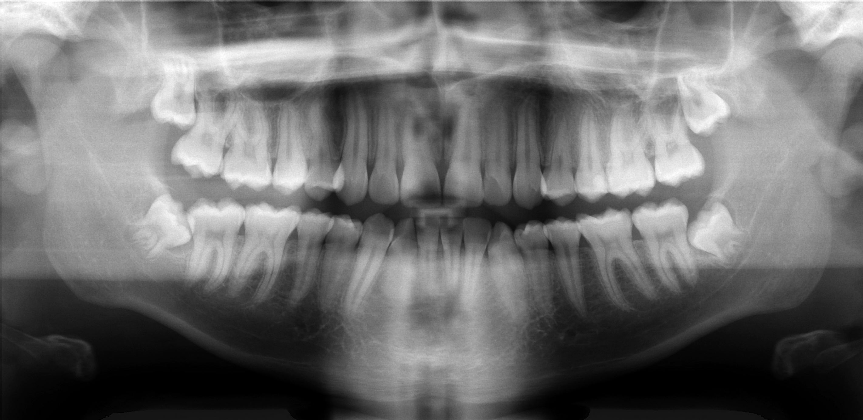
A dentist-validated guide to reading tooth numbers on a dental X-ray: the flipped left-right view, the two landmarks that orient you, and shape tips for telling each tooth apart.
Validated by our dental expert team Independent dentists providing online second opinions.
Your treatment plan says "crown, #30," and your X-ray is full of grey shapes that all look roughly the same. Matching the number on the page to the actual tooth on the film feels like it should be simple, and once you know the trick, it is. This guide shows you how to find any tooth number on a dental X-ray, with plain-English tips for telling the teeth apart by their shape, so a line on your plan finally points to a place you can see.
Quick Answer
On a US dental X-ray, teeth are labeled with the Universal Numbering System (1 to 32), and you find any tooth by combining two things: the fixed numbering path and the tooth's shape on the image. The numbering starts at the upper right wisdom tooth (number 1), runs across the top to the upper left wisdom tooth (16), drops to the lower left wisdom tooth (17), and runs back across the bottom to the lower right (32). The single most important trick is that an X-ray is flipped left to right, so the patient's right side appears on the left of the image. Find the midline between the two front teeth, then count back toward the molars, using root count and crown size to confirm each tooth. If you would rather have a dentist match every number to the image for you, that is exactly what an online dental second opinion does.
First, get the numbering path in your head
Before you can point to a tooth on a film, it helps to know the path the numbers follow. In the United States, dentists use the Universal Numbering System, which runs in one continuous loop: number 1 is the upper right wisdom tooth, the count sweeps across the top to number 16 (the upper left wisdom tooth), drops down to number 17 (the lower left wisdom tooth), and runs back across the bottom to number 32 (the lower right wisdom tooth). If that loop is new to you, read tooth numbering explained first, then come back here to apply it to an actual image.
The one rule that unlocks everything: the film is flipped
Here is the rule that trips up almost everyone. A dental X-ray is mounted and read as though you are looking at the patient face to face. That means the image is a mirror of your own mouth: the patient's right side appears on the left of the picture, and the patient's left side appears on the right.
So when you see the wisdom tooth at the far left of an upper film, that is tooth number 1 (upper right), not number 16. It feels backwards at first. The fix is simple: whenever you look at your own X-ray, remember that the left of the screen is your right, and the numbers on the top row will read 1 on the far left up to 16 on the far right.
Two landmarks that orient you instantly
You do not count all 32 teeth every time. You find one landmark and count out from it.
- The midline. Look for the gap between the two central front teeth. On the top, that gap sits between tooth 8 (the patient's upper right central incisor) and tooth 9 (the upper left central incisor). On the bottom, it sits between 24 and 25. The midline is your anchor for the front of the mouth.
- The last tooth in the corner. The tooth at the very back of each corner is the wisdom tooth (third molar): numbers 1, 16, 17, and 32. If a wisdom tooth has been removed, the second molar (2, 15, 18, or 31) becomes the last tooth. This is your anchor for the back of the mouth.
Pick whichever landmark is closest to the tooth you are trying to identify, then step tooth by tooth toward it.
Tell the teeth apart by shape (the part most guides skip)
The reason an X-ray looks like a wall of grey shapes is that no one showed you what each tooth type looks like. Once you know the shape cues, you can confirm a number instead of guessing. Reading from the front of the mouth to the back:
- Incisors (the four front teeth in each arch) have thin, single roots and small, flat crowns. Up top these are numbers 7, 8, 9, and 10; on the bottom, 23, 24, 25, and 26.
- Canines have the longest, stoutest single root in the whole mouth, and they sit right at the corner where the arch turns from front to side. A single very long root is almost always a canine (numbers 6, 11, 22, and 27).
- Premolars are smaller than molars, usually with a single root, and there are two of them between the canine and the molars. (The upper first premolar often shows two roots, which can briefly look molar-like, but its smaller crown gives it away.)
- Molars have the biggest crowns and multiple roots. On the image, upper molars usually show three roots and lower molars usually show two, spreading apart like legs. A big tooth at the back with more than one root is a molar (first molars 3, 14, 19, 30; second molars 2, 15, 18, 31).
One more shortcut: bright, solid white shapes on the film are metal fillings, crowns, posts, or root fillings, which block X-rays completely (they are described as radiopaque). If your plan mentions work on a tooth that already has a filling or crown, spotting that bright white restoration is often the fastest way to find the exact tooth. For more on reading the light and dark areas, see the dental X-ray interpretation guide for patients.
Start at the back: finding the molars and wisdom teeth
The back of the mouth is the easiest place to build confidence, because the molars are large and distinctive and the wisdom tooth marks the end of the count.
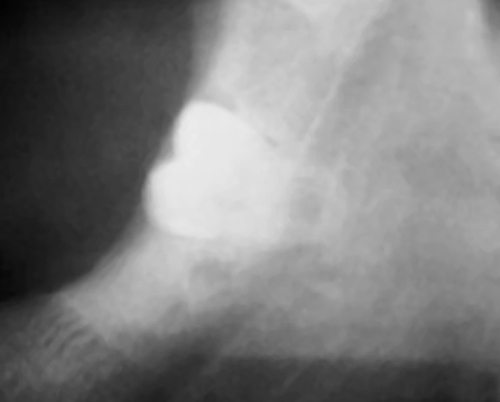
The tooth at the very end is the third molar, or wisdom tooth: number 1, 16, 17, or 32 depending on the corner. Wisdom teeth are also the teeth most likely to be sideways (impacted), partly erupted, or missing entirely, which is worth knowing because a missing or angled wisdom tooth can throw your count off if you are counting gaps instead of teeth. Count teeth, not spaces. Moving forward from the wisdom tooth, the next large multi-rooted teeth are the second molar, then the first molar, which is the single most commonly treated tooth in the mouth.
Walking a full-mouth (panoramic) X-ray, number by number
A panoramic X-ray shows both jaws and every tooth on one wide image, which makes it the best film for practicing the whole count.
Work the top arch first. Remembering the left-right flip, the tooth at the far left of the image is the upper right wisdom tooth (1), and the count climbs across the top: second molar (2), first molar (3), two premolars (4 and 5), the long-rooted canine (6), then the incisors (7 and 8) reaching the midline. Cross the midline and the numbers continue up the far side: 9 and 10 (incisors), 11 (canine), 12 and 13 (premolars), 14 and 15 (molars), and 16 (the upper left wisdom tooth) at the far right.
The bottom arch follows the same idea in reverse. Directly below the upper left wisdom tooth sits the lower left wisdom tooth (17), and the count runs back across the bottom to the lower right wisdom tooth (32), with the two lower central incisors (24 and 25) meeting at the midline. If you can walk a panoramic like this once, every smaller film becomes easier.
Bitewings and periapicals: numbering on close-up films
Most people are not handed a panoramic. The everyday films are smaller:
- Bitewings show the crowns of the upper and lower back teeth on one side, pressed together as if you are biting down. They are used mainly to find decay between teeth, so they usually show a run of premolars and molars from one side. Identify the side (left or right), find the most-forward premolar or the most-back molar, and count within that short run.
- Periapicals show a few whole teeth including the root tips. Because they are close-ups, you rely more on shape (Is this the long single root of a canine? A three-rooted upper molar?) and on any landmark in view, then match against your plan.
If your records came with written notes rather than the raw images, our guide to understanding your dental X-ray report and chart notes decodes the shorthand you will see alongside the numbers.
When the count shifts: missing teeth, extractions, and baby teeth
A few real-world situations change what you see:
- Missing or previously extracted teeth leave a gap, but the numbers do not renumber to close it. Tooth 30 is still tooth 30 even if 29 was pulled years ago. Count by position in the arch, not by the visible spaces.
- Teeth removed for braces. Some people had premolars extracted for orthodontics, which can make the arch look short. The molars are still 2 and 3 (or 14 and 15, and so on).
- Baby (primary) teeth use letters A through T instead of numbers, following the same loop around the mouth. A child's film labeled with letters is normal.
Why matching the number to the image matters
Being able to place each number is not trivia. It lets you take every line on your treatment plan and confirm it lands on the tooth you expect. If you remember a sensitive tooth on your lower left and the plan lists a crown on number 19, those line up. If the plan lists major work on a number you cannot find on your own film, that is a perfectly reasonable thing to ask your dentist to point out to you. To connect the numbers to the rest of the document, see how to read your dental treatment plan, and remember you have a right to copies of the images themselves under the guidance in how to get your dental records and X-rays. The American Dental Association maintains the standardized numbering and codes dentists use, and its consumer site MouthHealthy is a friendly place to read more about tooth types.
FAQ
How do I know which tooth is number 30 on my X-ray? Number 30 is your lower right first molar. On the flipped image it sits on the lower arch toward the right of the picture (which is your lower right). Find the back wisdom-tooth corner, count forward past the second molar, and the next large multi-rooted tooth is number 30, often the most heavily restored tooth on the film.
Why does the left side of my X-ray show my right teeth? Because dental films are mounted as though the dentist is facing you, the image is mirrored. The patient's right side appears on the viewer's left. Once you expect that flip, the top row reads number 1 on the far left up to number 16 on the far right.
How can I tell a molar from a premolar on the image? Look at size and roots. Molars have the largest crowns and multiple roots (upper molars usually three, lower molars usually two). Premolars are smaller and usually have a single root. A big tooth at the back with spreading roots is a molar.
What are the bright white shapes on my X-ray? Solid bright white areas are metal or dense materials such as fillings, crowns, posts, or root fillings, which block X-rays. They are useful landmarks: if your plan mentions a tooth that already has a restoration, the white shape helps you pinpoint it.
Do baby teeth have numbers on an X-ray? In the Universal system, the 20 primary (baby) teeth are labeled with letters A through T rather than numbers 1 to 32. The lettering follows the same path around the mouth, so a child's film with letters is completely normal.
Can I identify tooth numbers without any dental training? For most everyday films, yes. Using the numbering path, the left-right flip, the midline and back-tooth landmarks, and the shape cues, you can locate the teeth on your plan. If a film is unclear or you want certainty, a dentist can confirm each number for you in writing.
Final Advice
Identifying tooth numbers on an X-ray comes down to a short routine: remember the image is flipped, anchor yourself at the midline or the back corner, and use crown size and root count to confirm each tooth as you step along the arch. Do that once on a full-mouth film and the smaller everyday films stop looking like a wall of grey.
This guide was reviewed and validated by our dental expert team before publishing. If you would rather not decode it alone, upload your X-rays and treatment plan to toothcheck for a plain-English online dental second opinion, and a licensed dentist will match every number to the image and explain what the plan means, usually in under 72 hours. You can also start with a focused dental treatment plan review.
Image credits: Impacted wisdom tooth radiograph by Coronation Dental Specialty Group via Wikimedia Commons (CC BY-SA 3.0); panoramic radiograph by Coronation Dental Specialty Group via Wikimedia Commons (CC BY 3.0). Used as educational examples — they do not depict toothcheck patients.
Last medically reviewed: July 2026