What Does a Cavity Look Like on an X-Ray?
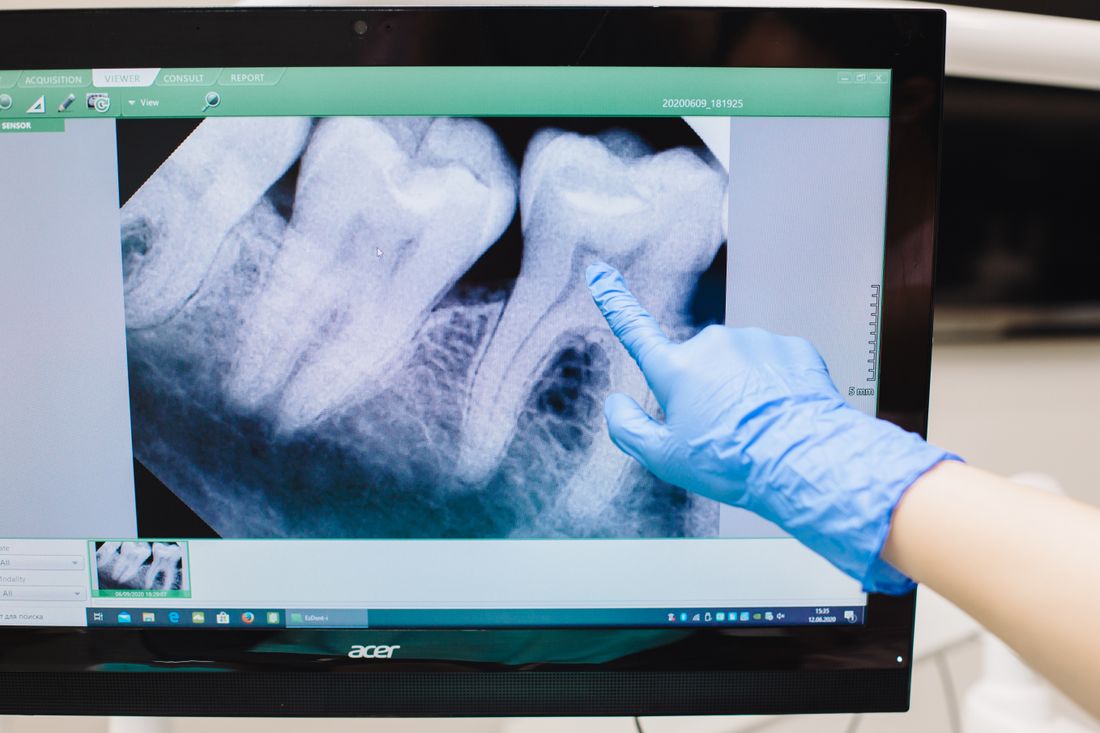
What does a cavity look like on an X-ray? Learn how decay shows as a dark shadow, how stages differ, and what X-rays can miss before you agree to a filling.
Reviewed by the toothcheck Dental Team Independent dentist providing online second opinions.
If your dentist has pointed at a grey blob on a screen and said "there is a cavity here", it can be hard to see what they see. Dental X-rays are shadow pictures, and learning to read even the basics makes it easier to ask good questions about whether a filling is needed.
This guide explains what a cavity looks like on an X-ray, why it looks that way, how the appearance changes as decay gets deeper, and, just as important, what X-rays cannot tell you. It is written to help you understand your dentist's explanation. It is not a way to diagnose yourself.
Quick Answer
On a dental X-ray, a cavity looks like a darker shadow inside a tooth that is otherwise light. Enamel and dentin are dense and absorb X-rays, so they appear light. Decay has lost mineral and absorbs less, so it appears darker. The easiest cavities to see are between teeth on a bitewing X-ray, where they often look like a dark notch or small triangle at the point where two teeth touch. How deep that shadow reaches, and whether it has crossed into the dentin, is what usually decides between watching it and filling it.
Why decay shows up as a dark area
An X-ray image is a map of how much radiation passes through each part of your mouth. Dense structures block more of the beam and look white or light grey. These are called radiopaque. Less dense structures let more through and look dark. These are called radiolucent.
Tooth enamel is the most mineralized tissue in the body, so it looks the brightest. Dentin, the layer beneath, is less mineralized and looks a slightly darker grey. The pulp chamber, which holds the nerve and blood vessels, is soft tissue and looks dark in the middle of the tooth.
Tooth decay is a process of acid dissolving mineral out of the tooth. The acid comes from bacteria feeding on sugars and other fermentable carbohydrates. As mineral leaves, that part of the tooth becomes less dense, and the X-ray records it as a darker patch. The NIDCR describes the basics of how decay develops at https://www.nidcr.nih.gov/.
Where cavities are easiest to see
Between the teeth: bitewing X-rays
A bitewing X-ray is taken while you bite down on a small tab, and it shows the crowns of the upper and lower back teeth together. It is the standard image for finding cavities between teeth, called interproximal cavities, because those spots are hidden from direct view and hard to reach with a probe.
On a bitewing, an early interproximal cavity usually appears as a small dark notch on the side of a tooth, just below the point where it touches its neighbor. As it progresses it often takes a triangular shape, with the wide base on the outer enamel and the point aiming toward the center of the tooth. When the triangle spreads sideways under the enamel surface, it can look like a dark wedge that has moved past the enamel-dentin junction.
On the biting surface
Cavities on the chewing surface, called occlusal cavities, are harder. The enamel there is thick, and the grooves and cusps overlap on a flat image. Decay can spread inside the dentin beneath intact-looking enamel without much change on the film. By the time it shows clearly as a dark band under the biting surface, it is often already well into the dentin. This is one of the main reasons a dentist also checks the tooth by sight and touch.
Under old fillings and near the gum
Decay can form at the edge of an existing filling or crown. On the X-ray it appears as a dark line or wedge hugging the filling, though this can be difficult to separate from the filling's own shape. Decay on the root surface, close to the gum line, shows as a dark scoop in the root and is more common as gums recede with age.
How the stages of decay look
Dentists often grade X-ray decay by how far it has gone. Different systems use slightly different labels, but a common pattern uses E for enamel and D for dentin. The table below is a simplified guide, not a diagnostic standard.
| Stage | Where the shadow reaches | How it tends to look | Typical approach |
|---|---|---|---|
| Early enamel (E1-E2) | Outer half or into inner half of the enamel only | Faint, small, soft-edged dark notch | Often monitored with fluoride and better cleaning; not an automatic filling |
| Into dentin (D1-D3) | Crosses the enamel-dentin junction and into the dentin | Clearer dark wedge or triangle, spreading sideways beneath the enamel | Usually a filling, especially as it nears the middle third of the dentin |
| Deep, near the pulp | Reaches close to the nerve chamber | Large dark area close to the dark pulp space | Larger restoration; may need a root canal if the nerve is involved |
Two caveats apply to this table. First, the labels are judgments about an image, and two dentists can grade the same shadow differently. Second, the stage on the film is only part of the decision. A small dentin lesion in someone with a low cavity risk may be treated differently from the same lesion in someone who gets new cavities every year. We cover that in more detail in our article on whether every cavity needs a filling: /blog/do-all-cavities-need-fillings
What X-rays can miss or get wrong
Understanding the limits of the image matters as much as understanding its appearance.
Early occlusal cavities can be invisible. The thick enamel and overlapping grooves can hide the early stages entirely.
Size is usually underestimated. A noticeable amount of mineral must be lost before the image changes, so the shadow on film is typically smaller than the true cavity. This is a well-known limitation of radiographs and is part of why dentists may find more decay than they expected once they open the tooth.
Not every dark area is decay. Several things can mimic a cavity:
- Cervical burnout, an optical effect near the neck of the tooth, where there is less tooth tissue in the beam path and the area looks darker even though it is healthy.
- Normal anatomy, such as the dark space where the pulp chamber sits, or overlap from a neighboring tooth.
- The edge of a filling or crown, which can create dark lines through contrast.
A single image is a single angle. If the beam is angled poorly, neighboring teeth overlap and hide or fake a cavity. This is why a repeat or different view is sometimes taken.
The picture is two-dimensional. A standard bitewing or periapical X-ray flattens a three-dimensional tooth. Toothcheck reviews 2D X-rays of this kind, not 3D CBCT scans. For the difference between image types, see /blog/which-dental-images-for-which-treatment
Because of all this, an X-ray alone never makes a diagnosis. The dentist combines it with a visual exam, a check for sensitivity or pain, your history of cavities, and sometimes photographs. If you want a broader introduction to what the different parts of an image show, our guide at /blog/reading-dental-xrays-what-you-can-see walks through it.
Questions worth asking your dentist
If a cavity has been flagged, these questions can help you understand the recommendation:
- Which tooth and which surface is it on?
- How far into the tooth does it appear to go: enamel only, or into the dentin?
- Can you show me on the X-ray, and is there any chance this is anatomy or burnout?
- How would you rate my risk of new cavities, and does that change the plan?
- Is it reasonable to monitor this one with fluoride and a recheck, and what would make you want to treat it?
- What happens if I wait six months?
A thoughtful answer to these tells you much more than the shadow alone does. It is also reasonable to ask for copies of your images, which many practices can provide in digital form.
FAQ
What does a cavity look like on an X-ray? A cavity usually appears as a darker area inside a tooth that is otherwise light grey or white. Decay removes mineral, so less X-ray energy is absorbed and the film or sensor records a darker shadow. Between back teeth it often looks like a small dark notch or triangle near where two teeth touch.
Can an X-ray miss a cavity? Yes. Early decay on the chewing surface can be hidden because the thick enamel and grooves overlap on the image. Small cavities may also not show until enough mineral has been lost to change the picture. A clinical exam is part of the diagnosis.
Is every dark spot on a dental X-ray a cavity? No. Normal anatomy, an old filling edge, and cervical burnout near the neck of the tooth can all look like decay. Dentists compare the X-ray with what they see and feel in the mouth rather than relying on one image.
Does a cavity that shows on an X-ray always need a filling? Not always. A shadow confined to the outer enamel is often monitored with fluoride and better cleaning, because enamel can remineralize. A shadow that has crossed into the dentin is more likely to need a filling. The right call depends on your cavity risk, the surface involved, and how the tooth looks clinically.
Why does my cavity look small on the X-ray but my dentist says it is bigger? X-rays tend to underestimate how deep and wide decay actually is, because a lot of mineral must be lost before the image changes. A cavity that looks modest on film can be larger once the dentist opens the tooth.
Can I diagnose my own cavity from my X-ray? No. This article is educational and is not a self-diagnosis tool. Only a licensed dentist who has examined you, or reviewed your images and records, can say whether a spot is decay and what to do about it.
Final Thoughts
A cavity on an X-ray is a dark shadow where a tooth has lost mineral, and the story it tells depends on where it sits and how deep it goes. The image is a useful clue, not a verdict, and it has well-known blind spots. For general guidance on cavities and prevention, the American Dental Association's patient site at https://www.mouthhealthy.org/ is a reliable starting point.
This article is for education only and does not replace an examination by a dentist. If you have been told you need a filling, or several, and want to know whether each one is needed now or can be watched, a written second opinion can tell you what your own X-ray shows and whether the plan fits it. You can get an online dental second opinion, where a licensed dentist reviews your own X-ray and tells you whether each filling is truly needed now or can be watched -- see also do all cavities need fillings?
Last medically reviewed: October 2026