What Does Bone Loss Look Like on a Dental X-Ray?
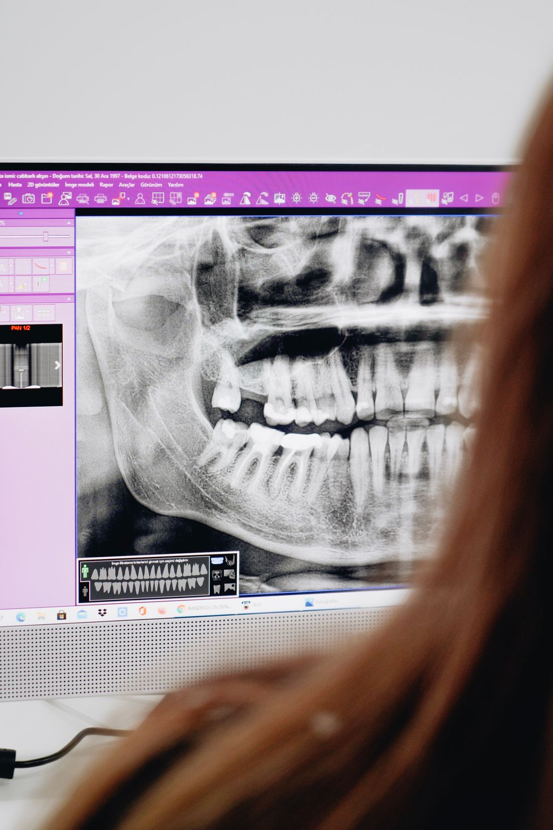
What does bone loss look like on a dental X-ray? Learn how dentists read bone levels, the patterns they look for, and what a film can and cannot show.
Reviewed by the toothcheck Dental Team Independent dentist providing online second opinions.
If your dentist has pointed to a dental X-ray and said "you have some bone loss," it can be hard to see what they mean. The film is a grey map of shadows, and the bone that matters is a thin line between the teeth. Knowing roughly what a dentist is looking at makes the conversation much easier to follow.
This guide explains what healthy bone looks like, how bone loss changes the picture, the common patterns dentists name, and the real limits of what an X-ray can tell you. It is meant to help you understand your records, not to diagnose yourself. Only a clinical examination combined with your films can do that.
Quick Answer
What does bone loss look like on a dental X-ray? The bone that supports the teeth appears as a grey-white structure around the roots. In health, its upper edge (the crest) sits about 1 to 2 mm below where the crowns meet the roots. With periodontal bone loss, the crest sits lower, further down the root, and the area between the teeth looks darker where bone used to be. The pattern can be even (horizontal) or a deeper notch beside one root (vertical or angular). An X-ray shows bone that has been lost, but not gum pocket depth or whether disease is active today.
What a healthy bone level looks like
On a bitewing or periapical film, each tooth has a crown covered in bright enamel and a root embedded in bone. Where the enamel ends and the root begins is called the cemento-enamel junction, or CEJ. Dentists use it as a landmark.
In a healthy mouth, the crest of the bone between two teeth sits roughly 1 to 2 mm below the CEJ on each neighbouring tooth. The crest usually looks like a fairly crisp, thin white line, and it often runs roughly parallel to an imaginary line joining the CEJs of the neighbouring teeth. The roots are surrounded by a thin dark line (the periodontal ligament space) and then a thin white line (the lamina dura, the bony socket wall).
What bone loss changes on the film
In periodontitis, the bacterial biofilm along the gumline triggers a chronic inflammatory response. Over time this breaks down the bone that holds the tooth in place. On the X-ray, you see:
- The crest of the bone sitting further from the CEJ, so more of the root is visible above the bone
- Dark (radiolucent) space where bone used to be, between the teeth
- A crest that looks less crisp or more irregular than in health
- In more advanced cases, roots that appear longer than the bone surrounding them, because less of the root is held in bone
Dentists often estimate severity by how much of the root length has lost its bony support. Rough, commonly used descriptions are mild, moderate, and severe, but the exact classification system used (for example the staging in the 2017 periodontal classification) depends on the dentist and combines X-ray findings with probing, attachment loss, and other factors.
Patterns dentists name
Horizontal bone loss
The bone crest recedes more or less evenly, so the bone level drops in a fairly flat line across several teeth. This is the most common pattern and is often described as the bone being "lower across the board."
Vertical (angular) bone defects
Here the bone has been lost more deeply along the side of one root than on its neighbour. On the film, this shows up as a notch or V-shaped dark area next to a single tooth. Vertical defects can be more localised, and the shape of the defect influences which treatments are considered.
Furcation involvement
Molars have two or three roots that split from a common trunk. The point where they divide is called the furcation. When bone loss reaches this area, you may see a dark shadow between the roots. This matters because the area is hard to clean and hard to treat. Furcation involvement is graded by how far into the split the loss extends, and it often needs a clinical probe (a special curved one) to assess properly.
A quick comparison
| Stage or pattern | What it can look like on the X-ray |
|---|---|
| Healthy | Bone crest about 1 to 2 mm below the CEJ, crisp thin line |
| Early or mild loss | Crest slightly lower than expected, sometimes subtle; may look slightly blunted |
| Moderate horizontal loss | Crest lower across several teeth, more root visible above bone |
| Severe loss | Bone around much of the root length is gone; roots look long |
| Vertical (angular) defect | Deep notch of missing bone beside one root |
| Furcation involvement | Dark shadow between the roots of a molar |
Which X-rays show it best
Different films show bone differently:
- Bitewings show the upper and lower back teeth together and are good for seeing the bone crest between teeth. Vertical bitewings, taken with the film oriented vertically, capture more of the bone height and are often used when bone loss is suspected.
- Periapical films show the whole tooth from crown to root tip and the surrounding bone, so they give the best view of how far down the root bone loss has gone.
- Panoramic films give an overview of all teeth and the jaws in one image. They are useful for spotting widespread patterns, but they are generally less precise for the fine bone detail around individual teeth.
For more on how these films compare, see our guide on which dental images for which treatment and our walkthrough of reading dental X-rays.
What an X-ray cannot tell you
This is where it is worth being careful. X-rays are valuable, but they are one piece of the picture.
- Pocket depth. The film shows bone, not the gum pocket around the tooth. Pocket depths are measured by gently probing with a small ruler-like instrument.
- Whether the disease is active. A film shows damage that has accumulated over months or years. It cannot tell you whether the gums are inflamed or whether bone is being lost right now. Bleeding on probing, changing pocket depths, and comparisons with earlier films help judge that.
- Early or slight loss. Small amounts of bone loss can be subtle or missed, particularly early on.
- Three-dimensional detail. A standard dental X-ray is two-dimensional. Bone on the cheek-side or tongue-side of a tooth can be hidden behind the root, so a film can under- or over-represent a defect. Overlapping teeth and the angle of the beam can also distort apparent bone height.
Because of these limits, a diagnosis of gum disease is made by combining the X-rays with a clinical examination, not from the X-ray alone. The American Dental Association (https://www.ada.org/) and the National Institute of Dental and Craniofacial Research (https://www.nidcr.nih.gov/) both describe periodontal disease as something diagnosed through a full evaluation.
If you have been told you have significant bone loss
Hearing that you have bone loss, and that you may need several deep cleanings, gum surgery, or tooth removal, can be unsettling. Some reasonable questions to ask:
- Which teeth are affected, and how severe is it at each one?
- What are my pocket depths, and are they consistent with what the X-rays show?
- Do you have older X-rays to compare, so we can see whether the loss is recent or long-standing?
- What happens if I treat only part of this, or wait?
- Are there alternatives to the plan you are recommending?
If the plan is extensive and you want a second look, a second dentist can review the same X-rays and the findings you have been given and say whether the plan seems consistent with the films. We cover what a gum disease recommendation should include in this guide. A second opinion works best when it is based on the actual films, not a description of them.
FAQ
Can an X-ray show gum disease? An X-ray shows the bone supporting the teeth, and bone loss is one consequence of periodontitis. It does not show gum inflammation, bleeding, or pocket depth, so diagnosis also needs a clinical exam with a probe.
What is a normal bone level on a dental X-ray? In a healthy mouth the bone crest usually sits about 1 to 2 mm below the cemento-enamel junction, where the crown meets the root. Bone sitting further down the root may indicate loss of support.
Does bone loss on an X-ray mean the disease is active right now? Not necessarily. A film shows what has been lost over time, not whether disease is progressing today. Probing, bleeding, and comparison with earlier X-rays help answer that.
Can lost bone grow back? Generally, bone lost to periodontitis does not regrow on its own. Treatment aims to stop further loss. In some specific defect shapes, regenerative procedures may be considered, and your dentist or periodontist can explain whether that applies to you.
Which X-ray is best for seeing bone loss? Bitewings, especially vertical ones, and periapical films show bone level around the teeth in the most detail. A panoramic film gives a useful overview but is generally less precise for fine bone detail.
I was told I need gum surgery based on my X-rays. Should I get a second opinion? It is reasonable to ask, especially for extensive treatment. A second dentist can review the same films and tell you whether the plan seems consistent with them.
Final Thoughts
Bone loss on a dental X-ray shows up as a lower bone crest, more visible root, and darker space between the teeth, sometimes as an even drop and sometimes as a deeper notch beside one root. It is a useful clue, but it is only part of a diagnosis. This article is educational and is not a diagnosis or a substitute for an examination by your own dentist. If you have been given a gum treatment plan and want a review of your films, you can request an online dental second opinion from an independent licensed dentist, typically within about 72 hours.
Last medically reviewed: October 2026