Types of Dental X-Rays Explained
A plain-English, illustrated guide to the types of dental X-rays - bitewing, periapical, panoramic, occlusal, cephalometric, and CBCT - what each shows and when it is taken.
Reviewed by the toothcheck Dental Team Independent dentist providing online second opinions.
If your dentist has ever said "let us take a couple of bitewings" or "we need a pan," you have probably nodded along without quite knowing what they meant - or why a particular X-ray was chosen. There are several distinct types of dental X-ray, and each one exists to answer a different question. Knowing which is which helps you understand your own diagnosis, follow your treatment plan, and recognise when an image is genuinely needed versus routine.
This guide walks through each type, in plain English, with a real example of what it looks like, and explains what it shows and when it is taken.
Quick Answer
Dental X-rays fall into two broad groups. Intraoral X-rays, where the film or sensor sits inside your mouth, include the bitewing (decay between back teeth and bone levels), the periapical (one or two whole teeth, from crown to root tip), and the full-mouth series (a complete set of those). Extraoral X-rays, taken from outside the mouth, include the panoramic (a single wide view of the whole jaw), the occlusal (a broad view of the arch), the cephalometric (a side view of the skull used in orthodontics), and CBCT (a 3D scan for complex cases). Which one you get should match the clinical question - and imaging should be based on your individual risk, not taken automatically.
Intraoral vs Extraoral X-Rays
The simplest way to organise dental imaging is by where the detector sits.
- Intraoral means the sensor is placed *inside* the mouth while the X-ray source points at it from outside the cheek. These give the most detailed, high-resolution views of individual teeth and are the workhorses of everyday dentistry.
- Extraoral means both the source and the detector are *outside* the mouth, usually on a machine that rotates around your head. These trade fine detail for a much wider field of view, capturing the whole jaw or skull.
The American Dental Association and the FDA share selection criteria for dental radiographs - the principle is that the *type and frequency* of imaging should be tailored to you, not applied on a fixed schedule to everyone.
Bitewing X-Rays (BW)
The bitewing is the most common dental X-ray, and the one most people have had many times without knowing its name. You bite down on a small tab (the "wing"), and a single image captures the *crowns* of the upper and lower back teeth together.
A bitewing is designed to answer two questions that the eye cannot: is there decay between the back teeth, in the tight contact points a mirror cannot see, and how are the bone levels around those teeth (an early sign of gum disease). It does not show the tips of the roots - that is a different view. Bitewings are the standard screening image at routine check-ups, and how often they are appropriate depends on your decay risk rather than a fixed calendar.
Periapical X-Rays (PA)
Where a bitewing shows only the crowns, a periapical shows one or two *entire* teeth - from the biting surface all the way down to the tip of the root and the bone surrounding it. "Peri-apical" literally means "around the tip."
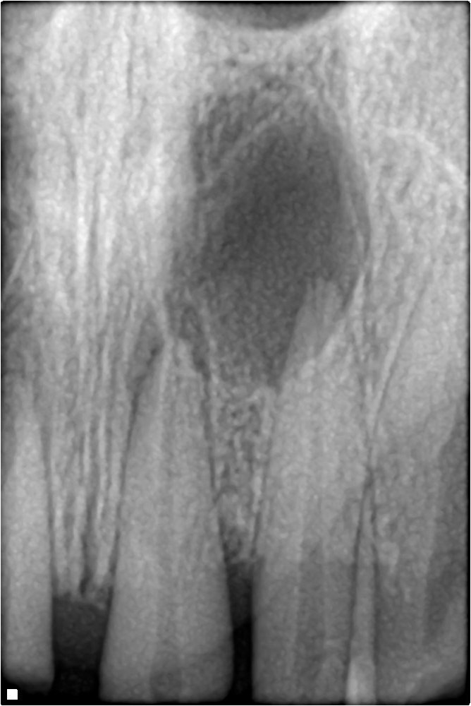
This is the view a dentist reaches for to investigate a *specific* problem: a toothache, a suspected abscess or infection at the root tip, a root fracture, or to check the roots before and after a root canal. If your dentist is focused on one tooth, you are usually getting a periapical. For help decoding what these findings mean, see our dental X-ray interpretation guide.
Full-Mouth Series (FMX)
A full-mouth series is not a different kind of image - it is a *complete set* of intraoral X-rays (a combination of periapicals and bitewings) that together document every tooth and its roots. It is typically taken for a new patient with a complex history, or periodically every few years, to give a comprehensive baseline. Because it involves many individual films, it is more involved than a couple of bitewings, and like all imaging it should be justified by your clinical situation rather than taken by default.
Occlusal X-Rays
An occlusal X-ray uses a larger film held between the teeth, giving a broad, flat view looking down onto (or up at) the dental arch - the roof or floor of the mouth. It captures a wider span of the jaw than a periapical.
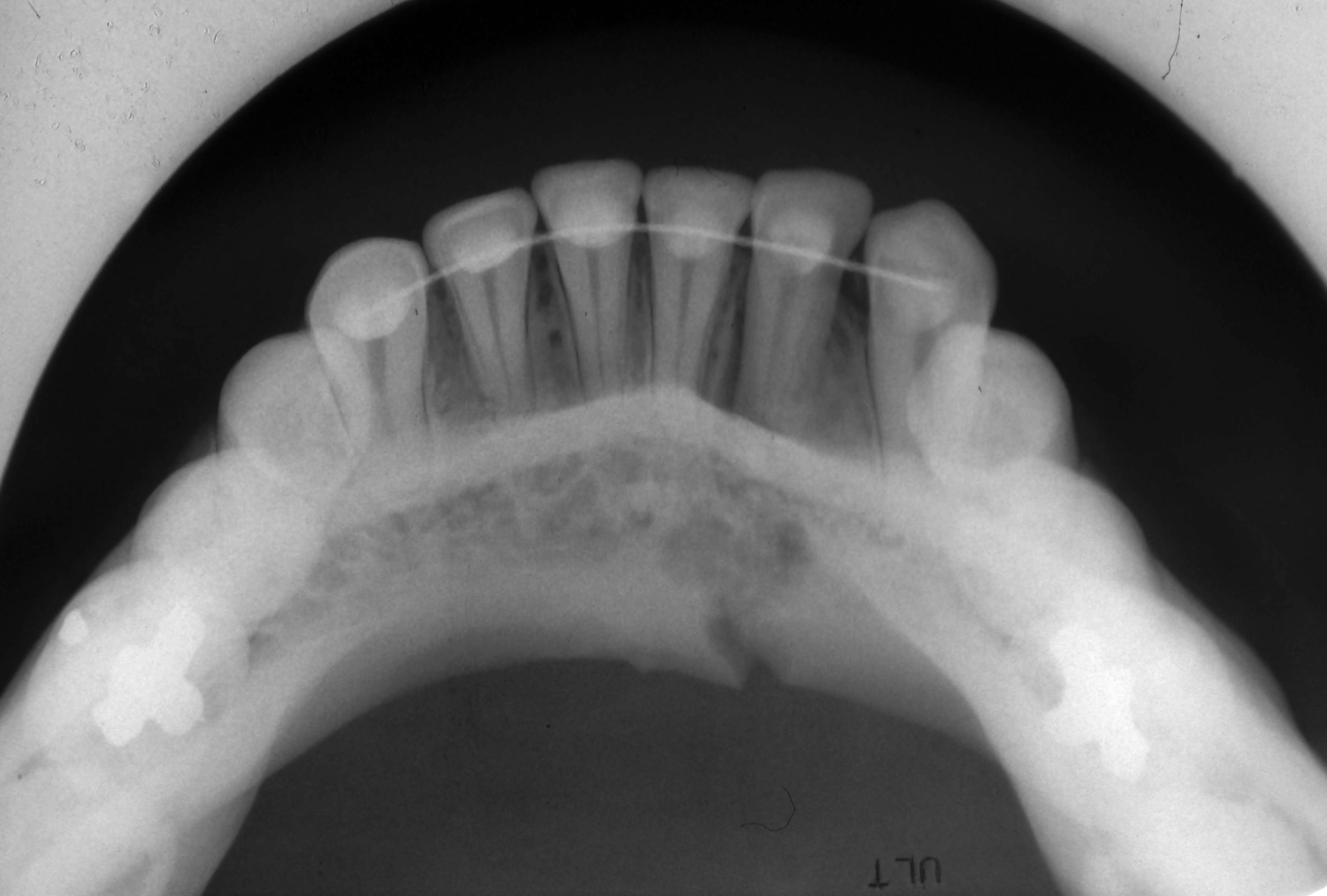
Occlusal views are used less often in routine dentistry now, but they remain useful for specific tasks: locating an unerupted or extra tooth, finding a stone in a salivary duct, assessing the extent of a cyst, or in children. If your care is routine, you may never need one.
Panoramic X-Rays (Pan / OPG)
The panoramic - often shortened to "pan" or called an OPG - is the sweeping, single image of your entire mouth: all the upper and lower teeth, both jaws, the sinuses, and the jaw joints, captured as the machine rotates around your head.
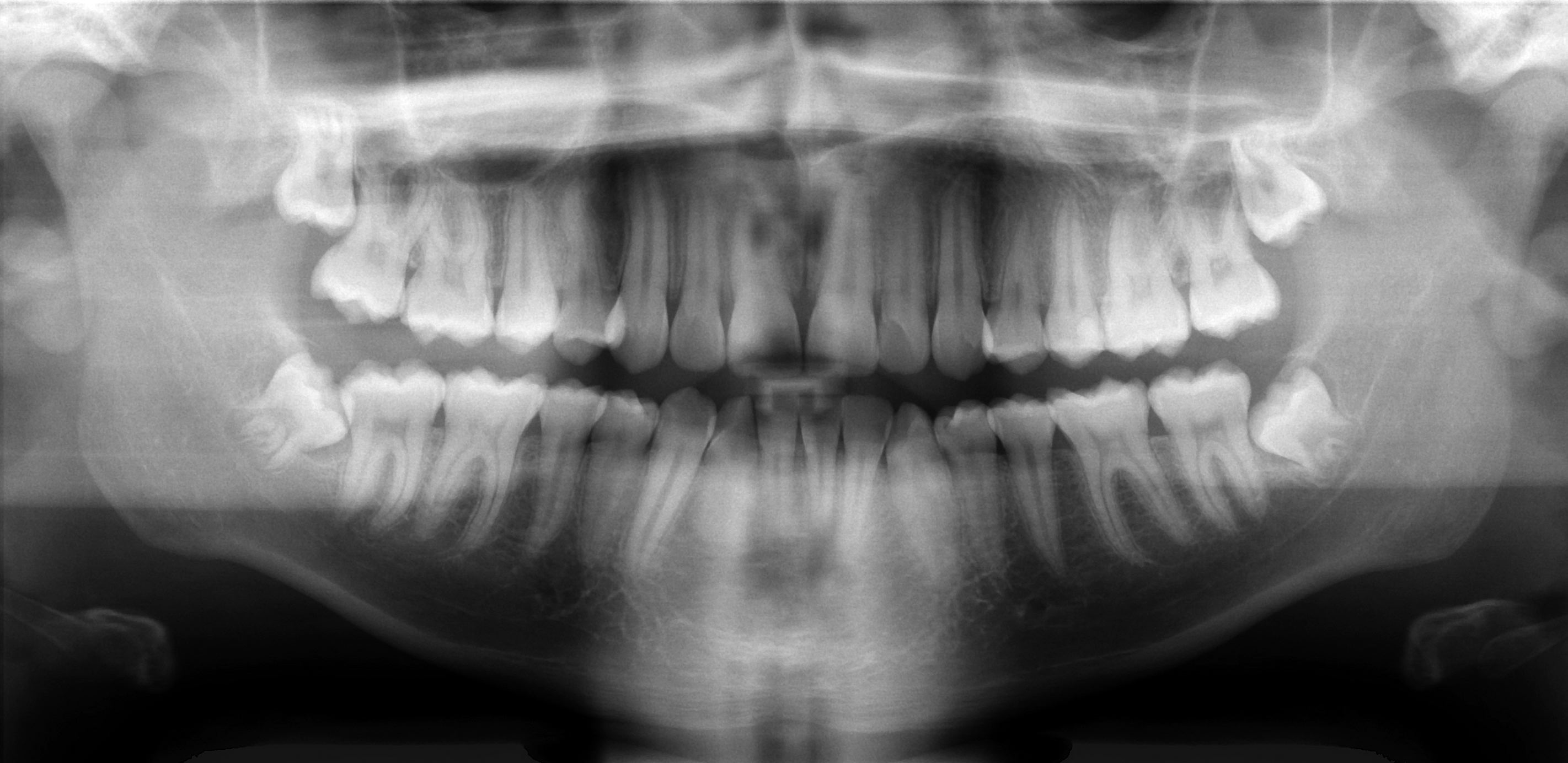
A panoramic is the go-to for the *big picture*: assessing wisdom teeth, planning extractions, screening for cysts or large lesions, checking jaw structure, and getting an overview for a new patient. Its trade-off is detail - it is excellent for scope but not as sharp as a bitewing for spotting a small cavity between two teeth, which is why the two are often used together.
Cephalometric X-Rays
A cephalometric (or "lateral ceph") is a side-on X-ray of the whole skull and jaws. It is used almost exclusively in orthodontics and jaw surgery.
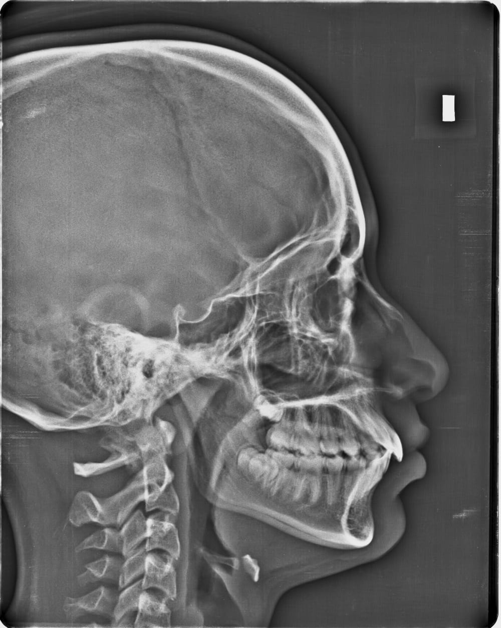
By showing the relationship between the teeth, the jaws, and the skull from the side, it lets an orthodontist measure growth and bite relationships and plan treatment. If you or your child are being assessed for braces, this is the specialist image you may encounter.
CBCT (Cone Beam CT) - 3D Imaging
CBCT, or cone beam computed tomography, is the most advanced dental imaging: a 3D scan built from many images taken as the machine rotates around your head, which can be viewed as cross-sections and reconstructions.
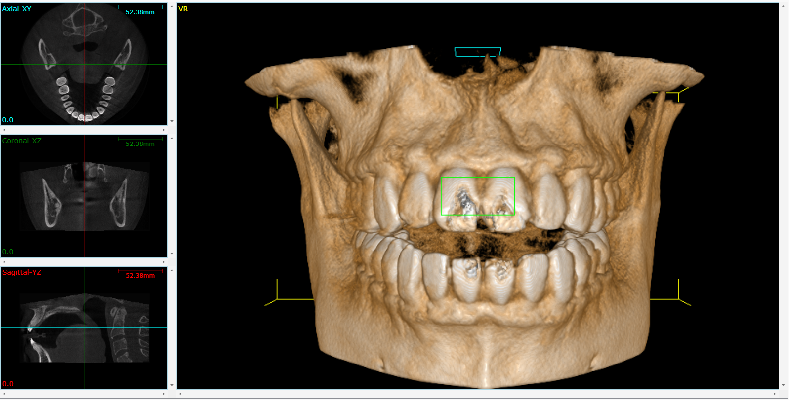
CBCT delivers far more information than a 2D X-ray - and a higher radiation dose - so it is reserved for situations that genuinely need it: implant planning (measuring bone height and the position of nerves and the sinus), complex root canals and suspected root fractures, impacted teeth close to a nerve, and jaw pathology. The American Academy of Oral and Maxillofacial Radiology publishes guidance on when CBCT is and is not indicated. A CBCT recommended for a routine problem that a standard X-ray could answer is worth a question - and if a large, imaging-heavy plan has you unsure, an independent second opinion can confirm what was actually needed.
Are Dental X-Rays Safe, and How Often?
Modern dental X-rays use very low doses of radiation, and digital sensors have reduced that further. The guiding principle across the profession is to image based on your individual risk and clinical need, not on a fixed timetable - which is exactly why the *type* chosen should map to a real question. If you want to understand the written findings that come back from any of these, see understanding your dental X-ray report and notes, and to connect imaging to what you are being charged, how to read your dental treatment plan.
FAQ
What is the difference between a bitewing and a periapical X-ray? A bitewing shows only the crowns of the upper and lower back teeth together and is best for finding decay between teeth and checking bone levels. A periapical shows one or two whole teeth from crown to root tip and is used to investigate a specific tooth and its root.
What is a panoramic X-ray used for? A panoramic captures the whole mouth in one wide image - all the teeth, both jaws, the sinuses, and the jaw joints. It is the standard view for wisdom teeth, extractions, and an overall assessment, though it is less detailed than a bitewing for small cavities.
Do I really need a CBCT (3D) scan? CBCT is reserved for complex situations - implant planning, difficult root canals, suspected fractures, impacted teeth near a nerve, or jaw pathology - where 2D imaging is not enough. It carries a higher radiation dose, so a CBCT for a routine problem a standard X-ray could answer is worth questioning.
Which dental X-ray is used for braces? A cephalometric X-ray, a side view of the skull and jaws, is used in orthodontics to measure the relationship between the teeth, jaws, and skull and to plan treatment. A panoramic is often taken alongside it.
How often should I have dental X-rays taken? There is no fixed schedule that fits everyone. The type and frequency should be based on your individual decay and gum-disease risk and your clinical situation, following ADA and FDA selection criteria - not taken automatically at every visit.
Are dental X-rays safe? Modern digital dental X-rays use very low doses of radiation. As with all imaging, they should be taken when there is a genuine clinical reason, which is why the right type is matched to the question being asked rather than taken by default.
Final Advice
Each dental X-ray exists to answer a specific question: a bitewing for decay between teeth, a periapical for a single tooth and its root, a panoramic for the whole jaw, a cephalometric for orthodontics, and CBCT for 3D planning when a flat image is not enough. Knowing which is which helps you follow your own diagnosis and recognise when an image genuinely fits the problem.
If you have been handed a set of X-rays and a treatment plan and you are not sure the imaging - or the treatment attached to it - was necessary, you can have it reviewed. Upload your X-rays to toothcheck for a plain-English independent second opinion from a licensed dentist, or start with a dental treatment plan review. You can also read our dedicated guide to getting a dental X-ray second opinion.
Image credits: Periapical radiolucency image by Shaimaa Abdellatif via Wikimedia Commons (CC BY-SA 4.0); occlusal radiograph by Coronation Dental Specialty Group via Wikimedia Commons (CC BY 3.0); panoramic radiograph by Coronation Dental Specialty Group via Wikimedia Commons (CC BY 3.0); lateral cephalogram by Challiyan via Wikimedia Commons (CC BY-SA 4.0); CBCT scan image by Panda 51 via Wikimedia Commons (CC BY-SA 4.0). Used as educational examples - they do not depict toothcheck patients.
Last medically reviewed: July 2026