What Does a Cracked Tooth Look Like on an X-Ray? (Why You Often Can't See It)
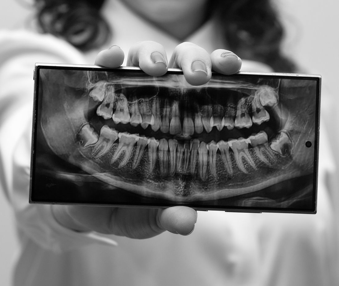
What does a cracked tooth look like on an X-ray? Often nothing at all. Learn why cracks hide, which indirect signs show up, and how cracks are really found.
Reviewed by the toothcheck Dental Team Independent dentist providing online second opinions.
Here is the honest answer first: very often, a cracked tooth looks like nothing at all on an X-ray. If you have been told your X-ray looks normal but your tooth still hurts when you bite, that is not a contradiction. It is one of the best-known limitations of dental imaging.
That is why this article spends as much time on what an X-ray cannot show as on what it can. If you came here hoping to spot a crack on your own image, the realistic expectation is that you probably will not, and neither will most people looking at a standard film. What you can learn is which indirect signs matter, how cracks are actually found, and what questions to ask about a plan.
Quick Answer
A cracked tooth on an X-ray is often invisible, because most cracks run in a plane roughly parallel to the X-ray beam and leave no shadow to see. When a crack has progressed, the X-ray may show indirect signs instead, such as a dark area at the root tip, a narrow vertical area of bone loss beside one root, or a visibly separated fragment. Cracks are mainly diagnosed in the chair, using your symptoms, a bite test, a light shone through the tooth, and sometimes dye. A normal X-ray never rules a crack out.
Why Cracks Hide on a Standard X-Ray
A dental X-ray is a shadow picture. The beam passes through the tooth and the sensor records how much was absorbed along the way. To see a line, the beam has to cross a gap wide enough, and aimed in the right direction, to let more radiation through along that line.
Most cracks do not meet those conditions. A typical crack in a back tooth runs from the biting surface down toward the root, often front to back (mesial to distal). The X-ray beam also travels from the cheek side toward the tongue side, so it runs along the crack, not across it. A hairline crack that is tightly closed and parallel to the beam adds almost no contrast. The image is also a flat, two-dimensional picture of a three-dimensional tooth, so structures overlap and a thin line is easily lost.
This is the reason a dentist can hold a perfectly clear X-ray in one hand and a patient who flinches at every bite in the other. The two are not in conflict. The image is simply not very sensitive for this question.
Authoritative sources describe the same limitation. The American Association of Endodontists and the ADA both note that cracks are frequently difficult to detect and that X-rays often do not reveal them (see https://www.ada.org/ for the ADA's patient resources on cracked teeth).
The Types of Cracks, and Why the Type Matters
Not every crack means the same thing. The names below are the commonly used categories, and the difference between them drives treatment.
- Craze lines: Tiny, shallow lines in the enamel only. Very common in adult teeth, painless, harmless, and not visible on X-rays. Usually no treatment is needed.
- Fractured cusp: A piece of the chewing surface has broken off or is about to, often around a large old filling. It tends to be painful on chewing but is generally not deep enough to reach the nerve, and it is usually restored with a filling or a crown.
- Cracked tooth: A crack that runs from the biting surface toward the root, with the tooth still in one piece. Symptoms vary, and the nerve may be affected. Treatment depends on how deep the crack goes, and often involves a crown, sometimes with a root canal.
- Split tooth: A crack that has progressed until the tooth separates into distinct segments. Usually the tooth cannot be saved in one piece.
- Vertical root fracture: A crack that begins in the root and extends upward. It can be quiet for a long time and may only be recognized when infection or bone loss appears. These often require extraction.
Why does this matter on an X-ray? Because the higher up the list, the less likely an image is to show anything. The lower down the list, the more likely there are secondary signs.
What an X-Ray Might Show: Indirect Signs
When a crack has been present long enough to cause damage, the body reacts to it, and that reaction can sometimes be seen. These are clues, and none of them is specific to cracks.
- A periapical radiolucency: A darker area at the tip of a root, which suggests infection or inflammation in the bone. It can be caused by deep decay or a failing nerve just as easily as by a crack.
- A vertical or J-shaped bone defect: A narrow channel of bone loss running alongside one root. When this occurs on a single tooth, particularly one with a root canal or a crown, it raises suspicion of a vertical root fracture. Gum disease can produce bone loss too, but typically in a more generalized pattern.
- A widened periodontal ligament space: The thin dark line around a root appears thicker, which can reflect inflammation or stress on the tooth.
- A visibly separated fragment: In a split tooth or a fractured cusp, a gap or displaced piece may be visible.
- A halo around a root: In some root fractures, bone loss outlines the root along its length.
The caveat is that by the time these signs appear, the problem is usually advanced. An X-ray is better at confirming damage than at catching an early crack.
How Cracked Teeth Are Actually Diagnosed
Since the film often cannot answer the question, clinicians rely on a combination of methods. The table below summarizes the main ones and what each can and cannot do.
| Method | What it involves | What it can tell you | Main limit |
|---|---|---|---|
| Symptom history | Questions about sharp pain on biting, pain on release, cold sensitivity | Strong clue about which tooth and what type | Pain can be vague and can refer to neighbouring teeth |
| Bite test | Biting on a small stick or rubber tool, one cusp at a time | Pain on release is a classic sign of a crack | Needs a cooperative patient; can be negative |
| Transillumination | A bright light shone through the tooth | A crack can block the light, appearing as a dark line | Does not show crack depth |
| Dye (such as methylene blue) | Stain painted on the tooth to highlight lines | Makes the crack path visible | Shows surface lines, not depth |
| Removing an old filling or crown | Taking out the restoration to inspect the tooth underneath | Allows direct sight of the crack | An invasive step; done only when warranted |
| Gum probing | Measuring pocket depth around the tooth | A narrow, deep pocket on one side suggests a root fracture | Not specific on its own |
| X-ray or 3D imaging | Image of the tooth and surrounding bone | Shows secondary signs and ruled-out other causes | Often misses the crack itself |
Put together, these steps let a dentist build a case. Rarely does any single test settle it. The ADA and the NIDCR (https://www.nidcr.nih.gov/) both describe cracked teeth as a diagnosis that depends on a thorough exam as well as imaging.
Symptoms That Should Prompt an Exam
You cannot diagnose a crack from symptoms alone, but certain patterns are worth reporting to a dentist promptly:
- A sharp, short pain when you bite down, especially pain that hits when you release the bite
- Pain that appears with certain foods, such as a seed or hard crust, and then settles
- Sensitivity to cold that is sharp and brief
- Pain that comes and goes in a way that is hard to pin to one tooth
- A pimple-like bump on the gum near one tooth
- A tooth that has a large filling or has had a root canal and is now tender to biting
None of these confirms a crack. Cavities, gum problems, a failing filling, and a bite that is too high can feel similar, and that is another reason an in-person exam matters.
Where an Online Second Opinion Helps, and Where It Does Not
We want to be direct about this. An online review of an X-ray by a licensed dentist can look at the image you send and note anything visible, including the indirect signs above. What it cannot do is confirm that a crack exists, rule one out, or tell you how deep it goes. Those answers come from a clinical exam. Toothcheck reviews 2D X-rays and photos, not 3D scans, and the images are not a substitute for being examined.
Where a second opinion can genuinely help is the decision that follows. If a dentist has told you a tooth is cracked and recommended a crown, a root canal, or an extraction, you may want a fresh view on whether the plan is proportionate to what the images show. Our guides on crowns versus fillings and root canal versus extraction explain the trade-offs in plain English. If you want to see what an X-ray review involves, you can get an online dental second opinion.
A sensible way to use a second opinion for a suspected crack is to treat it as a way to understand the plan and prepare questions, while still getting the tooth examined in person. Useful questions include: how was the crack confirmed, how deep does it appear to go, is the nerve involved, what is the realistic chance of keeping the tooth, and what happens if we wait.
Treatment Depends on the Type, Not the Word Cracked
Because the word crack covers everything from harmless craze lines to an unsaveable root fracture, the plan should match the type. A fractured cusp is often restored. A cracked tooth with a healthy nerve may be protected with a crown. If the nerve is irritated or infected, a root canal followed by a crown is a common route. When a split tooth or vertical root fracture is confirmed, extraction is the usual outcome, because the crack provides a path for bacteria that cannot be sealed.
A note of caution applies in both directions. Treating a tooth aggressively on the strength of an unconfirmed crack is one risk. Delaying treatment of a true crack is another, because a crack that deepens can turn a restorable tooth into one that cannot be saved. This is exactly the kind of decision where a clinical exam and, if you wish, a second professional view are worth having.
FAQ
Can a dentist see a cracked tooth on an X-ray? Sometimes, but often not. Most cracks run in a plane roughly parallel to the X-ray beam, so they cast no visible shadow. A crack that has widened, split the tooth, or caused infection or bone loss around the root is more likely to show, usually through its side effects rather than the crack line itself.
If my X-ray looks normal, does that mean my tooth is not cracked? No. A normal X-ray does not rule out a crack. If you have sharp pain on biting, pain when you release a bite, or lingering cold sensitivity, a clinical exam is what answers the question: a bite test, a light shone through the tooth, dye, and a close look at the tooth and gum.
What are craze lines, and are they a problem? Craze lines are very fine, shallow lines in the enamel that most adult teeth develop over time. They are generally harmless and cosmetic, and they do not show on X-rays. They are different from a crack that runs deeper into the tooth and causes symptoms.
What on an X-ray might suggest a cracked tooth? Indirect signs: a dark area at the tip of a root, a narrow vertical or J-shaped area of bone loss along one side of a root, a widened gap around the root, or a visibly separated fragment. These are not specific to cracks and can have other causes, so they are clues that call for an exam, not a diagnosis.
Can an online second opinion diagnose a cracked tooth from an X-ray? No, and we say so plainly. A review of an X-ray alone cannot confirm or exclude a crack. It can point out indirect signs and help you understand and weigh a proposed plan, but a suspected crack needs an in-person clinical exam.
Do cracked teeth always need to be pulled? No. It depends on the type and depth of the crack. Craze lines need nothing, a fractured cusp is often restored with a filling or crown, and a cracked tooth may be saved with a crown, with or without a root canal. Split teeth and vertical root fractures usually cannot be saved.
Final Thoughts
If there is one thing to take from this article, it is that a normal-looking X-ray does not mean a tooth is intact. Cracks are mainly found by listening to your symptoms and examining the tooth, and the film is best seen as supporting evidence. If biting hurts, get the tooth examined in person rather than waiting for something to show up on an image.
This article is educational and is not a diagnosis or a substitute for an in-person examination by a dentist. If you have been given a treatment plan for a cracked tooth and want an independent view of it, you can learn how our online dental second opinion works. For general information about tooth fractures and dental care, see the ADA at https://www.ada.org/ and the NIDCR at https://www.nidcr.nih.gov/.
Last medically reviewed: October 2026