What Does an Impacted Wisdom Tooth Look Like on an X-Ray?
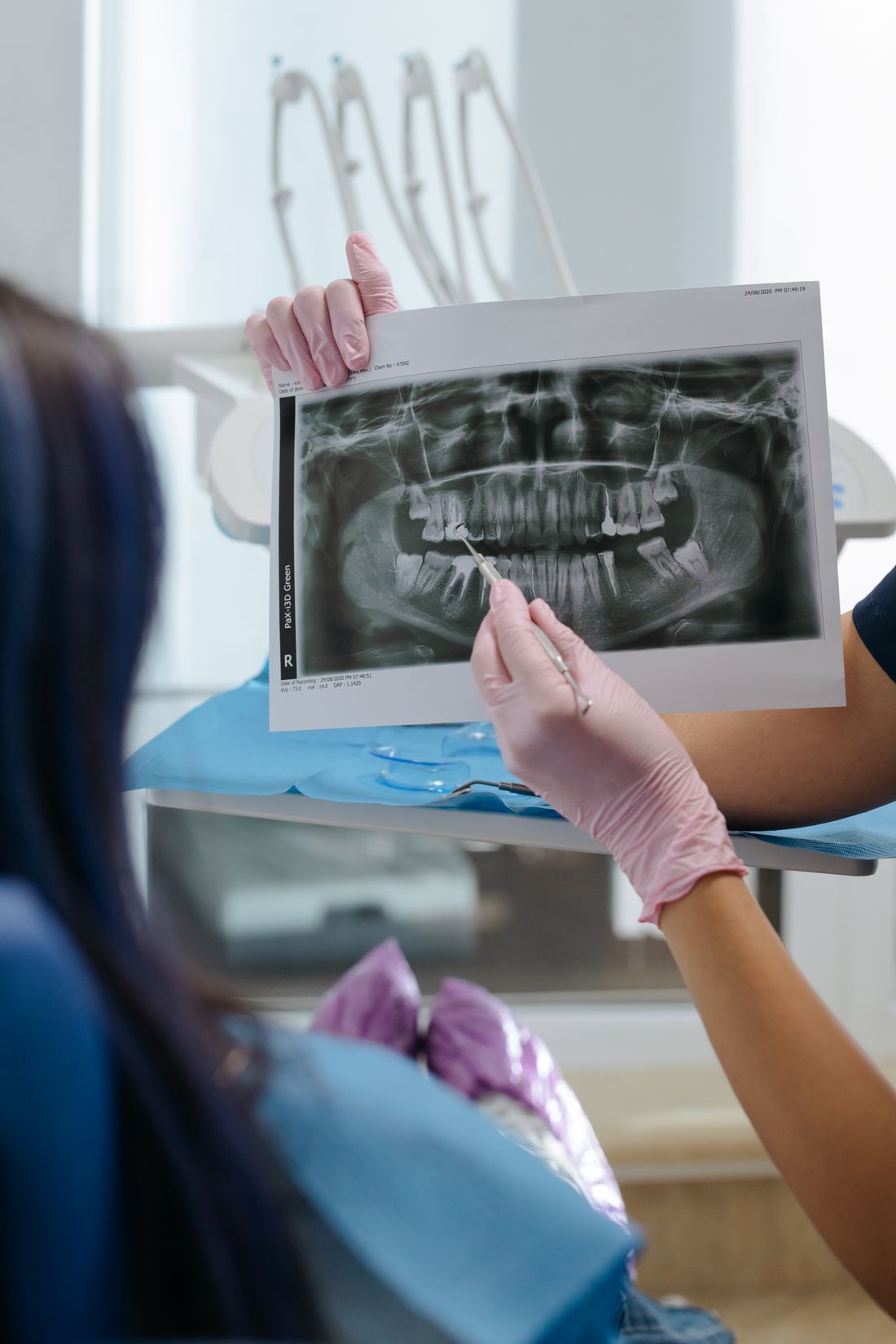
What does an impacted wisdom tooth look like on an X-ray? Learn the angles, bone cover, nerve proximity and warning signs a panoramic image can show.
Reviewed by the toothcheck Dental Team Independent dentist providing online second opinions.
If you have been told you have an impacted wisdom tooth, the first thing a dentist does is show you an X-ray, usually a wide curved image of both jaws. To the untrained eye it is a grey, ghostly picture of teeth and bone, and it is hard to see what the dentist is pointing at. This article explains what that image shows, in plain terms.
An impacted wisdom tooth (a third molar) is one that cannot fully come through the gum into its normal position, because it is blocked by the tooth in front, by bone, or by the angle it is growing at. The X-ray is how dentists judge the angle, how deeply the tooth is buried, and whether it is pressing on anything important. It is educational background only: it cannot replace an examination, and you should not diagnose yourself from an image.
Quick Answer
On an X-ray, an impacted wisdom tooth looks like a third molar sitting at the back of the jaw that is tilted, sideways or buried rather than standing upright like the other molars. The most common pattern is a tooth tilted forward toward the second molar (mesial impaction). Dentists also look at how much bone covers the tooth, whether it touches the second molar, whether the dark rim around the crown looks enlarged, and how close the roots of a lower wisdom tooth are to the nerve canal. An impacted tooth is not automatically a problem that needs removal.
Which X-Ray Is Used?
The standard view for wisdom teeth is the panoramic X-ray (sometimes called an OPG or orthopantomogram). The machine rotates around your head and produces one wide image showing all the teeth, both jaw joints and the nerve canal in the lower jaw. Because it captures the very back of the mouth, it shows third molars better than small bitewing films, which usually stop at the second molar.
Smaller periapical films of a single area are sometimes used for detail. The limitation of all these 2D images is that they flatten a three-dimensional jaw into a single picture, so structures at different depths overlap. For a general explanation of the image types, see our guide to which dental images are used for which treatment.
Reading the Angle: Types of Impaction
The angle of the tooth is the first thing described in a report. It is judged by comparing the long axis of the wisdom tooth with the long axis of the second molar in front of it.
| Type | What it looks like on the image |
|---|---|
| Mesial | Crown tilted forward, leaning into the back of the second molar. Generally the most common pattern. |
| Vertical | Tooth points the right way but is stuck, usually under bone or gum. |
| Distal | Crown tilted backward, away from the second molar, toward the back of the jaw. |
| Horizontal | Tooth lies on its side, with the crown pointing at the second molar's root. |
You may also hear terms such as angular or inverted, and positions described in relation to the jaw bone and the chewing surface of the neighbouring tooth. These classification systems are used by dentists and surgeons to estimate how technically involved a removal might be, not simply whether the tooth is a problem.
Soft Tissue Versus Bony Impaction
Impactions are also described by what covers the tooth. In a soft-tissue impaction, the crown is mostly through the bone but still partly covered by gum. In a partial bony impaction, part of the crown is covered by bone. In a complete bony impaction, the tooth is fully enclosed in bone.
On an X-ray, bone shows as a lighter (more radiopaque) layer over the crown, while the gum is not clearly visible. This is one reason the clinical examination matters: the image shows bone cover well but cannot reliably show whether a tooth is partly covered by gum. Depth matters in practice because teeth buried in bone usually need bone removed during surgery, while teeth only under gum may be simpler to access.
Pressure on the Second Molar
One of the most useful things the X-ray shows is the contact between the wisdom tooth and the second molar. Mesial and horizontal teeth can press against the back of the second molar and create a tight pocket that is hard to clean. Over time this can contribute to decay on the back surface of the second molar, or, less commonly, to resorption, where the root or crown of the neighbouring tooth is gradually worn away.
On the image, decay behind the second molar can appear as a darker notch in the enamel where the wisdom tooth touches it. Because the second molar is a tooth most people want to keep, this finding often carries weight in the decision about the wisdom tooth. It is also something to ask about specifically if you are weighing up removal. Small areas of early decay can be hard to see on a 2D image, and not every shadow is decay.
The Dark Rim Around the Crown
Each developing tooth is surrounded by a thin sac called the follicle, which shows on X-rays as a narrow dark (radiolucent) rim around the crown. A thin rim is normal.
A rim that looks clearly enlarged can sometimes indicate a cyst, most often a dentigerous cyst, which forms around the crown of an unerupted tooth. Most are slow-growing, but they can enlarge over time and occasionally damage nearby bone. Whether a dark area is a normal follicle or something that needs attention is a professional judgement, often involving comparison with the other side and follow-up imaging. The X-ray alone cannot establish what a lesion is, and the NIDCR (https://www.nidcr.nih.gov/) provides patient information on wisdom teeth and related problems.
Nerve Proximity in the Lower Jaw
The roots of lower wisdom teeth can lie close to the inferior alveolar nerve, which runs inside a canal in the lower jaw and supplies feeling to the lower lip and chin. On a panoramic image, the canal appears as a thin dark band with fine white borders running below the tooth roots.
Dentists look for signs that suggest a closer relationship, including:
- Darkening of the root where the canal crosses it
- Narrowing of the root
- Interruption of the white lines of the canal wall
- A canal that appears to bend or divert near the root
These signs raise the possibility of closer contact, though they are not proof. Because a panoramic image is 2D, the canal can seem to overlap the root while actually running to the inside or outside of it. When the question matters for surgical planning, a 3D cone beam CT (CBCT) scan is often ordered in person to see the true position. toothcheck reviews 2D X-rays and does not interpret CBCT, so nerve-proximity cases sometimes need that in-person step. Altered feeling after wisdom tooth surgery is uncommon, and surgeons weigh the risk for each case.
Do All Impacted Wisdom Teeth Need to Come Out?
No, and this is a genuinely debated area. Removal is generally advised when there is a clear problem: repeated gum infection around a partly covered tooth (pericoronitis), decay in the wisdom tooth or the second molar, gum disease around the area, a cyst, or damage to the neighbouring tooth. The ADA (https://www.ada.org/) lists wisdom teeth among topics where treatment should be based on the individual patient's situation.
For impacted teeth with no symptoms and no disease on examination or X-ray, some clinicians recommend monitoring with periodic review, while others favour earlier removal because surgery is often easier at a younger age. Neither view is automatically right. Reasons to lean toward monitoring include a deeply buried tooth with no contact problems, higher surgical risk, or a tooth close to the nerve. Reasons to lean toward removal include difficulty keeping the area clean, early decay or a tooth you cannot realistically keep healthy.
If you are weighing this up, it is reasonable to ask which specific finding on your X-ray is driving the recommendation. Our guides on wisdom tooth extraction second opinions and alternatives to tooth extraction go into the decision in more detail.
Questions Worth Asking Your Dentist
- Which angle and depth is my wisdom tooth, and does that change how difficult removal would be?
- Is there contact or decay on the second molar?
- Is the dark rim around the crown normal in size?
- How close are the roots to the nerve, and do I need a 3D scan?
- What happens if we monitor instead of removing it, and what would we watch for?
FAQ
Can you tell if a wisdom tooth is impacted from an X-ray? Usually yes. A panoramic X-ray shows the third molars, their angle, bone cover and contact with the second molar. A clinical exam of the gum is still needed to judge whether the tooth is partly covered by soft tissue.
What is the most common angle of an impacted wisdom tooth? Mesial impaction, tilted forward toward the second molar, is generally described as the most common. Vertical, distal and horizontal impactions are the other main types.
Does every impacted wisdom tooth have to come out? No. Impacted teeth with no symptoms and no disease are sometimes monitored. Whether to remove them preventively is genuinely debated, so it is reasonable to discuss it with your dentist or surgeon.
How can an X-ray show that a wisdom tooth is close to the nerve? Signs include roots overlapping the nerve canal, darkened or narrowed roots, or a canal that seems to bend near the root. A 2D image cannot show the true 3D relationship, so a 3D scan is often ordered in person when this matters.
What is the dark area around some impacted wisdom teeth? A thin dark rim is the normal follicle. A clearly enlarged rim can occasionally indicate a cyst and needs professional assessment. The X-ray alone does not give a diagnosis.
Can toothcheck review my panoramic X-ray for wisdom teeth? Yes. A licensed dentist can review a 2D panoramic X-ray and the plan you were given. We do not interpret 3D CBCT scans.
Final Thoughts
An impacted wisdom tooth on an X-ray is a matter of angle, depth, contact and distance from the nerve, and none of these findings on their own decides whether a tooth must be removed. This article is educational and is not a diagnosis or a substitute for an examination by your own dentist or surgeon.
If extraction of one or more wisdom teeth has been recommended and you are unsure, an online dental second opinion from a licensed dentist can review your panoramic X-ray and the proposed plan for 59 dollars, with a written response in about 72 hours.
Last medically reviewed: October 2026