Which Dental X-Rays Matter for Which Treatment (and What to Send)
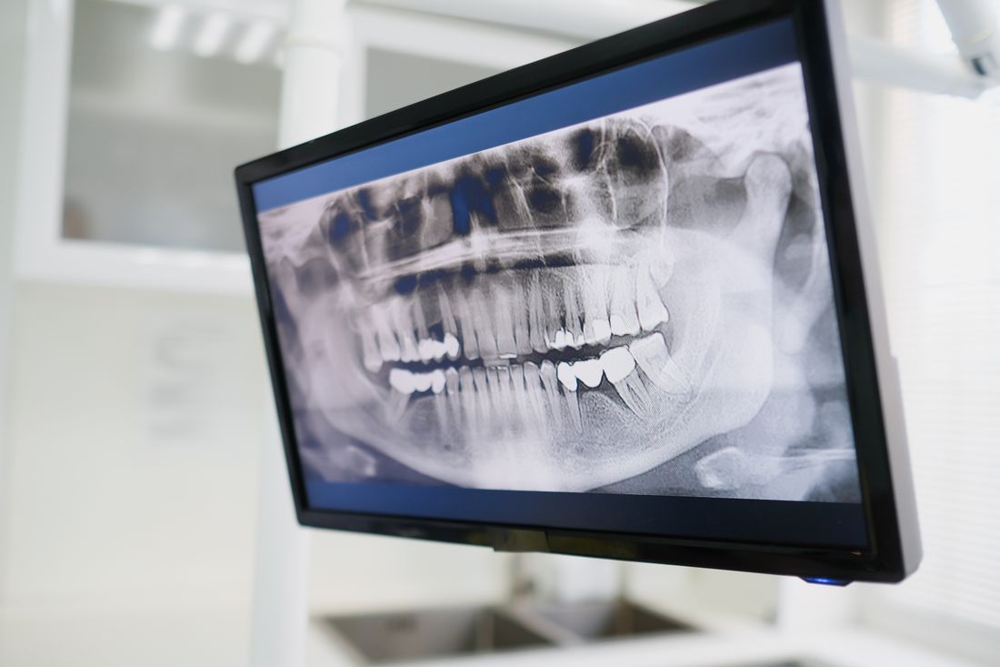
Which dental X-rays matter for which treatment? Bitewings, periapicals, panoramics and photos: what each shows, what it misses, and what to send.
Reviewed by the toothcheck Dental Team Independent dentist providing online second opinions.
Dental X-rays are not interchangeable. Each type is designed to answer a particular question, and a treatment plan can only be judged properly if the right image is in front of the reviewer. A set of bitewings can tell you a lot about whether back-tooth cavities are real, yet almost nothing about whether a tooth with a toothache needs a root canal. A panoramic image can show a wisdom tooth well, but it is too blurry to confirm a small cavity between two molars.
This matters when you are asking for a second opinion. A common reason a review is slower or less conclusive than it could be is that the images sent do not match the question being asked. This guide maps each image type to the treatment decisions it can and cannot support, so you know what to request from your dentist and what to send.
Quick Answer
Which dental X-rays matter for which treatment depends on the question. Bitewings answer "do I have cavities between my back teeth, and do these fillings need replacing." Periapical X-rays answer "does this tooth need a root canal or extraction." A panoramic X-ray answers full-mouth questions such as wisdom teeth, widespread bone loss and large rehabilitation plans. Close-up photos cover cracks, wear and chewing-surface decay that films can miss. For most cavity, filling, crown and root canal questions, bitewings plus periapicals, the written plan and one photo are enough for an online dental second opinion.
The Image Types at a Glance
| Image type | What it shows | Treatment question it answers |
|---|---|---|
| Bitewing X-ray | The crowns of upper and lower back teeth together, the spaces between them, and the bone crest | Is there decay between back teeth? Is there new decay under an old filling or crown edge? Do these fillings really need doing? |
| Periapical X-ray | One to three whole teeth from crown to root tip, with the surrounding bone | Does this tooth need a root canal, a retreatment or extraction? Is there an abscess? Is the root long and solid enough to restore? |
| Panoramic X-ray (OPG) | All teeth, both jaws, jaw joints and sinuses in one wide image | Wisdom teeth, generalized bone loss, missing teeth, full-mouth extraction, denture or full-arch implant plans |
| Intraoral photos | The visible surfaces: cracks, chips, wear, gum colour and swelling, staining, cavitated decay | Is this crack serious? Is this wear or erosion? Does the chewing-surface cavity actually exist? Do the gums look inflamed? |
| Written treatment plan | Tooth numbers, procedures and fees proposed | Is every item necessary, in a sensible order, and fairly priced? |
Bitewing X-Rays: The Cavity and Filling Question
A bitewing is taken while you bite down on a small tab, so the crowns of the upper and lower back teeth appear together. Because the X-ray beam passes through the contact points between neighbouring teeth, bitewings are the best routine image for decay between back teeth, which is called interproximal decay. This is the spot where cavities most often start and where they are hardest to see with the eye or a probe.
Bitewings are also how a reviewer checks the edges of existing fillings and crowns for new decay underneath. If your question is "do I really have all of these cavities" or "do these old fillings really need replacing," bitewings are usually the main evidence.
They also show how deep a lesion has gone. Decay confined to the outer enamel is a different situation from decay that has reached the inner layer, and that difference is often what separates "watch and re-image" from "fill now." For more on that distinction, see do all cavities need fillings.
What bitewings do not reliably show
Honesty about limits is important here, because bitewings are sometimes treated as if they show everything.
- The biting (occlusal) surface. Decay in the grooves of a chewing surface can be well advanced before it appears on a bitewing, because the dense enamel on top hides it. Occlusal decay often needs a clinical exam and a good photo.
- Roots. Root-surface decay near the gumline, especially where the gums have receded, is only partly visible.
- Root tips. A bitewing is framed on the crowns and the top of the bone. The ends of the roots are usually outside the picture.
- Everything about a lesion. X-rays tend to underestimate how deep decay actually is. Some lesions only reveal their true extent once the dentist starts treatment. A reviewer can say that a filling looks reasonable or questionable from the image, but cannot promise what will be found under the drill.
Periapical X-Rays: The Root Canal vs Extraction Question
A periapical film ("PA") is aimed at a specific area and captures the entire tooth from the crown down past the root tip into the surrounding bone. That is exactly the information needed for questions about the nerve and the root.
A tooth whose nerve has died, or a tooth with an infection at the tip of the root, often shows a dark area around the root end. Reviewers call this a periapical radiolucency. A PA also shows root shape and length, existing root canal fillings and whether they reach the tip, the width of the ligament space around the root, fractures that run along the root in some cases, and how much healthy bone is left.
For a decision such as root canal vs extraction, the PA is the key image. Without it, a reviewer can comment on the written plan, but cannot confirm whether the tooth is likely to be saveable. If a dentist is recommending a root canal, a crown after a root canal, or removal of a particular tooth, ask for a recent periapical of that tooth.
A caution: a dark area does not always mean a current infection, and a normal-looking root end does not always mean a healthy nerve. The X-ray is read together with symptoms, tests such as cold and tapping responses, and the clinical exam. A reviewer will say what the image supports and what it cannot settle remotely.
Panoramic X-Rays: The Whole-Mouth Question
A panoramic image (also called an OPG or pan) rotates around your head and produces one wide picture of all teeth, both jaws, the jaw joints and part of the sinuses. It exposes you to relatively little radiation for the area covered, and it is the right image when a decision involves the entire mouth.
It is most useful for:
- Wisdom teeth: their position, how they sit against neighbouring teeth and how close they are to the main nerve canal in the lower jaw.
- Generalized bone loss: an overall impression of how much bone supports the teeth, relevant to gum disease and to whether teeth can realistically be kept.
- Large plans: a recommendation to remove many or all teeth, make dentures, or place full-arch implants cannot be properly judged without the whole-mouth view. In our reviewed cases, several big plans simply could not be assessed responsibly until a panoramic was added.
- Missing and retained teeth, jaw cysts and similar findings that sit outside the area a small film covers.
The trade-off is detail. A panoramic image is lower resolution than bitewings or periapicals and distorts in places, so it should not be used alone to diagnose small cavities or fine root details.
Intraoral Photos: What Films Miss
Some of the most important findings are visible rather than radiographic. A clear photo, taken in good light with your phone, adds information that an X-ray cannot:
- cracks and craze lines, chips and broken cusps
- tooth wear and erosion, such as flattened biting surfaces or thinned front teeth
- gum colour, swelling and recession
- staining, white spots and visible decay on chewing surfaces
Photos help most when paired with X-rays. A bitewing may look clean while a photo of the chewing surface shows a cavitated groove, or a photo may show a crack in a tooth whose film looks normal. Take photos of the problem tooth from the top and from the side, with the cheek held back and no flash glare.
The Written Treatment Plan: The Piece People Forget
The images show the tooth. The plan shows what is being recommended. A reviewer needs the written plan with tooth numbers and the procedures proposed against each, ideally with the fees. Without it, we can describe your X-rays but not judge whether the plan is necessary, whether the sequence is sensible, or whether the price is in a normal range. A photo of the printed plan or a screenshot of the patient portal works fine. See dental treatment plan review for what that involves.
The Exception: When 3D Is Genuinely Needed
Some questions cannot be answered with flat images, and it is better to say so plainly. A cone-beam CT (CBCT) is a 3D scan taken in person at a practice or imaging centre. It can be the only way to answer questions such as:
- the exact position of certain impacted teeth, for instance buried canines or wisdom teeth very close to a nerve
- bone height, width and nerve and sinus position before placing implants
- suspected root resorption, where the root is being slowly dissolved from the inside or outside
- some complex root anatomy or suspected fractures
toothcheck does not interpret CBCT scans, so please do not upload one. If your situation falls into one of these categories, the review will tell you that a 3D scan is warranted and that you should see a specialist in person. That is a more honest answer than guessing from a 2D image. Most routine cavity, filling, crown and root canal questions do not need 3D imaging at all.
What to Send, by Situation
| If your question is | Send |
|---|---|
| Do I really have these cavities or need these fillings? | Bitewings, one photo of any tooth the plan names, the written plan |
| Do I need a root canal or extraction on one tooth? | A recent periapical of that tooth, a bitewing, the written plan |
| A crown is recommended | Bitewing and periapical of the tooth, a photo, the plan |
| Wisdom teeth | Panoramic, plus the plan |
| Many extractions, dentures or full-mouth implants | Panoramic, all available bitewings and periapicals, photos of the whole mouth, the plan |
| Cracked, worn or sensitive teeth | Clear photos, any recent X-rays, the plan |
For the practical steps, see how to upload dental X-rays for a second opinion. To learn what normal and abnormal findings look like on each film, see reading dental X-rays: what you can see. Our dental X-ray review page explains how the review itself works.
Two practical points. First, ask for the original digital files rather than photographing a screen if you can, since a phone picture of a monitor loses detail. Second, recency counts: images from several years ago may not reflect the mouth today. For general information on dental X-ray safety and use, the American Dental Association publishes patient guidance at https://www.ada.org/.
FAQ
Which X-ray do I need to check whether I really have cavities? Bitewing X-rays are the standard image for decay between back teeth and under existing fillings. They are less reliable for decay on the biting surface or on roots, so a clinical exam and close-up photos matter too.
What X-ray shows whether I need a root canal or an extraction? A periapical X-ray. It shows the whole tooth from crown to root tip plus the surrounding bone, which is where an abscess or a dead nerve leaves a visible dark area. A bitewing usually cuts off the root tip, so it cannot answer this question on its own.
When is a panoramic X-ray necessary? When the decision involves the whole mouth: wisdom teeth, widespread bone loss, extracting many teeth, dentures or full-mouth implants. A plan of that size is hard to judge without seeing all the teeth and both jaws in one image.
Do I need a 3D CBCT scan to get a second opinion from toothcheck? No, and please do not upload one. We review 2D X-rays, photos and written plans. Some situations, such as certain impacted teeth, implant planning or suspected root resorption, genuinely need a 3D scan done in person. If your case is one of them, we will tell you.
What if I only have a few images, or they are old? Send what you have, and ask your dental office for the rest. Recent images matter most because decay and bone change over time. If the images are not enough to judge your plan, the review will say exactly what is missing rather than guess.
Do I have to send photos as well as X-rays? It is strongly recommended. Cracks, wear, gum condition and decay on the chewing surface often show up better in a clear photo than on a film. A phone photo taken in good light is enough.
Final Takeaway
Match the image to the question. Bitewings for cavities and fillings, periapicals for a specific tooth and its root, a panoramic for whole-mouth decisions, photos for what films cannot see, and always the written plan. Be wary of any recommendation that rests on an image that cannot show the thing being treated, and know that a few questions truly need an in-person 3D scan. A licensed dentist reviews your X-rays, photos and plan for a flat 59 dollars, with a written answer in about 72 hours. Start your online dental second opinion.
Last medically reviewed: October 2026