Reading a Dental X-Ray: What You Can See at Each Stage of Decay and Disease
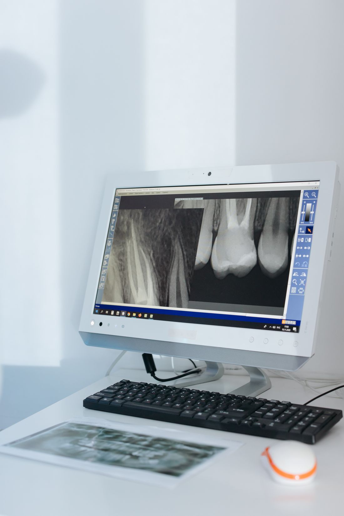
Reading a dental X-ray: what early decay, deep cavities, abscesses and gum bone loss look like, and what an X-ray cannot show. Educational, not diagnostic.
Reviewed by the toothcheck Dental Team Independent dentist providing online second opinions.
Most people are handed a treatment plan that says "cavity on number 14" or "bone loss in the lower back" and never see what the dentist is looking at. Yet the X-ray is the single most important document behind many of those recommendations, and the logic of reading it is simpler than it looks. Once you know that dense things are white and damaged things are dark, a lot of the picture starts to make sense.
This guide walks through what a dental X-ray can show at each stage of tooth decay and gum disease, what it cannot show, and why those limits matter. It is educational. Reading an X-ray properly takes years of training and almost always needs a clinical exam alongside it, so please treat this as a way to understand your images and ask better questions, not as a way to diagnose yourself.
Quick Answer
Reading a dental X-ray comes down to density. Enamel, fillings and crowns are dense and appear white, while decay, infection and bone loss are less dense and appear darker. Decay is staged by how far the dark shadow reaches: enamel only, into the dentin, near the nerve, or beyond the root tip. Gum disease shows up as bone that has dropped away from the crowns. X-rays are powerful but incomplete, so a dentist combines them with a clinical exam.
The Basic Rule: White Is Dense, Dark Is Damaged
An X-ray is a shadow picture. The beam passes through your mouth and the sensor records how much arrives on the other side. Dense material absorbs more of the beam and leaves the sensor lighter. Dentists call this radiopaque. Less dense material lets more of the beam through and appears darker, which is called radiolucent.
In practice that gives you a rough map:
- Brightest white: metal fillings, crowns, implants and root canal filling material.
- Light grey-white: enamel, which is the hardest tissue in the body, forming a bright outline around each crown.
- Mid grey: dentin, the softer layer underneath enamel, and healthy bone.
- Dark: the pulp chamber and root canals (soft tissue inside the tooth), the periodontal ligament space around each root (a thin dark line), and any area that has lost mineral.
Decay is simply tooth that has lost mineral to acid, so it looks like a darker patch cutting into a brighter structure. Shape and location help a dentist tell decay from normal anatomy, from an old restoration, or from an imaging artifact.
Which X-Ray Shows What
Different views are good at different things, which is a big reason a single image can look unhelpful. A bitewing, taken while you bite on a small tab, shows the crowns of upper and lower back teeth together with the bone level between them. It is the main tool for finding cavities between teeth and early bone loss. A periapical image captures a whole tooth from crown to root tip with the surrounding bone, and is what you look at to assess the root and what lies beyond it. A panoramic image sweeps around the entire jaw, which is useful for wisdom teeth, large cysts and an overview, but is less detailed for small cavities. If you are unsure which of these you need to share, see our guide on which dental images matter for which treatment.
Stages of Decay on an X-Ray
Decay does not appear all at once. It advances through layers, and the layer it has reached largely decides the approach.
Stage 1: early decay in the enamel. A faint shadow, often triangular and sitting just below the contact point where two teeth touch, that stays within the enamel and has not reached the dentin. Enamel at this point has lost some mineral but the surface may still be intact. Many dentists will monitor these, often with fluoride, better cleaning between the teeth and dietary changes, because early lesions can remineralize and stabilize. It is not an automatic filling. Whether to wait depends on your age, how many new cavities you have had, and how the dentist judges the rate of progression over time.
Stage 2: decay into the dentin. Here the shadow crosses the junction between enamel and dentin and spreads wider underneath, often in a wedge or a broader, flatter pattern. Dentin is softer than enamel, so decay moves faster once it gets there. A lesion that is clearly into the dentin is usually restored with a filling, though the timing is still a judgment call for a lesion that has only just crossed over. If you are unsure whether yours really needs treatment, our article on whether all cavities need fillings goes into this boundary in detail.
Stage 3: deep decay near the pulp. The dark area now reaches deep into the dentin and approaches the pulp chamber, where the nerve and blood vessels live. At this point the main question is whether the nerve is still healthy enough to be protected. A dentist may try to remove the decay and place a protective liner or a filling, or the nerve may be too inflamed or infected and a root canal becomes the likely path. The X-ray cannot tell you which one applies. Symptoms such as lingering pain to hot or cold or spontaneous night-time aching, along with clinical tests, do much of that work.
Stage 4: periapical change at the root tip. When infection from a dead or dying nerve spreads out of the root, bone around the tip can break down. On the image this appears as a dark area around the end of the root, called a periapical radiolucency. It generally points to an infected or non-vital tooth, and the options are usually a root canal or extraction. Some of these areas are small, long-standing and symptom-free, while others are linked to swelling or pain. Not every dark area at a root tip is an abscess, and a tooth that has been root-treated may take months to show bone healing. For examples of how a root canal recommendation is judged, see do I really need a root canal.
| Stage | How it looks on the X-ray | Typical approach |
|---|---|---|
| Early (enamel only) | Faint shadow within the enamel, not past the enamel-dentin junction | Often monitored; fluoride, hygiene and diet changes |
| Into the dentin | Clearer dark wedge or patch past the enamel-dentin junction | Usually a filling |
| Deep, near the pulp | Dark area approaching the pulp chamber | Deep filling or protective liner; root canal if the nerve is involved |
| Periapical change | Dark area around the root tip | Root canal or extraction, confirmed by clinical tests |
Gum Disease on an X-Ray
Periodontal disease is different from decay because the problem is the supporting bone, not the tooth. In a healthy mouth the bone sits a small distance below the crowns, and the ridge between neighbouring teeth looks like a crisp peak. In periodontitis the bone around the teeth is gradually lost because of chronic infection and inflammation.
Two patterns are common. Horizontal bone loss means the whole level drops evenly, so the crests of bone sit lower along the root. Angular (or vertical) loss means a V-shaped defect beside one tooth, which often points to a more localized problem. Dentists grade how severe the disease looks partly by how much of the root length has lost its bone: early, moderate or advanced. Calculus, the hardened deposit under the gum, can also sometimes be seen as small bright spurs on the root surface.
The limit is important. An X-ray shows where bone is now, but not whether the gums are bleeding, how deep the pockets are, or whether the disease is active. Those findings come from probing and a clinical exam, which is why a periodontal diagnosis needs both. If a treatment plan includes deep cleaning or gum treatment, check that your X-ray and your probing measurements agree.
Other Things a Dentist Looks For
Beyond decay and bone, the same images show a lot of useful detail. Existing fillings and crowns can be checked for gaps at the edge, overhangs that trap plaque and irritate the gum, or new decay creeping in around the margin. Previous root canals can be examined for how completely the canal was filled and whether the tip looks healthy. Retained root fragments, impacted or unerupted teeth, some cysts and sinus relationships may also be visible. These are the findings that often explain a recommendation to replace a restoration that looks perfectly fine in the mouth.
What an X-Ray Cannot Show
This is the section that matters most for avoiding both over- and under-treatment, because the gaps in an X-ray are where judgment comes in.
- Very early decay on the biting surface. The grooves of the chewing surface lie in the path of the beam alongside thick enamel, so a cavity starting there can be missed until it is well advanced. Dentists find these by looking, probing gently and sometimes using other tools.
- Hairline cracks. Cracks are usually too thin to show and are found by symptoms, bite testing, magnification and transillumination.
- Whether a nerve is alive or dead. The image may suggest a problem, but vitality is tested clinically with cold, pressure and percussion.
- The source of a particular pain. A perfect-looking X-ray does not mean nothing is wrong, and a dramatic-looking one may be completely painless.
- Soft tissue problems. Gum inflammation, mouth sores and many lesions of soft tissue are invisible.
- The full three-dimensional picture. A standard X-ray flattens a 3D object into two dimensions, so overlapping teeth and structures can hide or mimic problems. Some cases, such as complex implant planning or hard-to-locate infections, call for a 3D scan, which we do not interpret at toothcheck. We review 2D X-rays and photos.
Image quality matters too. A slightly angled bitewing can make overlapped teeth look decayed when they are not, and a periapical that cuts off the root tip simply cannot answer the question being asked.
Why Your Own X-Ray Is Worth Understanding
A treatment plan is a set of conclusions drawn from images, an exam and a conversation. When you understand roughly what the images show, you can ask questions that actually help: how deep is this shadow, is it into the dentin, is it being watched or treated, what makes you think the nerve is involved, and how does this connect to my symptoms. Good dentists welcome those questions. If you want to take your images for review, our guide on how to upload dental X-rays for a second opinion explains what to gather.
For general background on cavities and gum disease, the American Dental Association (https://www.ada.org/) and the National Institute of Dental and Craniofacial Research (https://www.nidcr.nih.gov/) publish reliable patient information.
FAQ
Why do cavities look dark on a dental X-ray? X-rays pass through less dense material more easily, so those areas look darker. Enamel, fillings and crowns are dense and look white. Decay has lost mineral, so it is less dense and shows as a darker shadow. The same logic applies to infection and bone loss.
Can I tell from my own X-ray if I need a filling? Not reliably. You may be able to spot a dark area, but whether it needs treatment depends on its depth, whether it is progressing, your cavity risk, and a clinical exam of the surface. A shadow limited to the enamel is often monitored, while one that has crossed into dentin usually is not.
What does a dark spot at the tip of a root mean? It is called a periapical radiolucency and often means bone has been lost around the tip because the nerve has died or become infected. It is not proof by itself, so a dentist confirms it with clinical tests and the history of the tooth.
Does an X-ray show gum disease? It shows the level of the bone between your teeth. Bone that has dropped away from the crowns suggests periodontitis. It does not show pocket depth, bleeding or inflammation, which need probing in the mouth.
What cannot be seen on a regular dental X-ray? Very early biting-surface decay, hairline cracks, whether a nerve is alive, the exact source of pain and soft-tissue problems are hard or impossible to see. A standard X-ray is also 2D, so overlapping structures can hide things.
Can a second opinion explain my X-ray to me? Yes. A licensed dentist can review your X-rays and photos and explain in plain English what they show and what each finding might mean for treatment. It supports your decisions and does not replace an in-person exam.
Final Thoughts
An X-ray is a map of density: white for solid structure, dark for places where mineral or bone has been lost. Decay is staged by how deep the shadow runs, from enamel through dentin to the nerve and beyond the root tip, and each stage points to a different approach. Gum disease is read from the bone level, while many important problems, such as cracks, nerve vitality and early biting-surface decay, need more than a picture.
Understanding this does not make you your own dentist, and it should not. What it can do is turn a confusing plan into one you can discuss. If you would like to know what your own X-ray actually shows, a written online dental second opinion from a licensed dentist can explain it in plain English, with a flat fee of 59 dollars and a typical turnaround of about 72 hours. You can also see how dental X-ray review works before you decide.
Last medically reviewed: October 2026