What Does a Dental Cyst Look Like on an X-Ray?
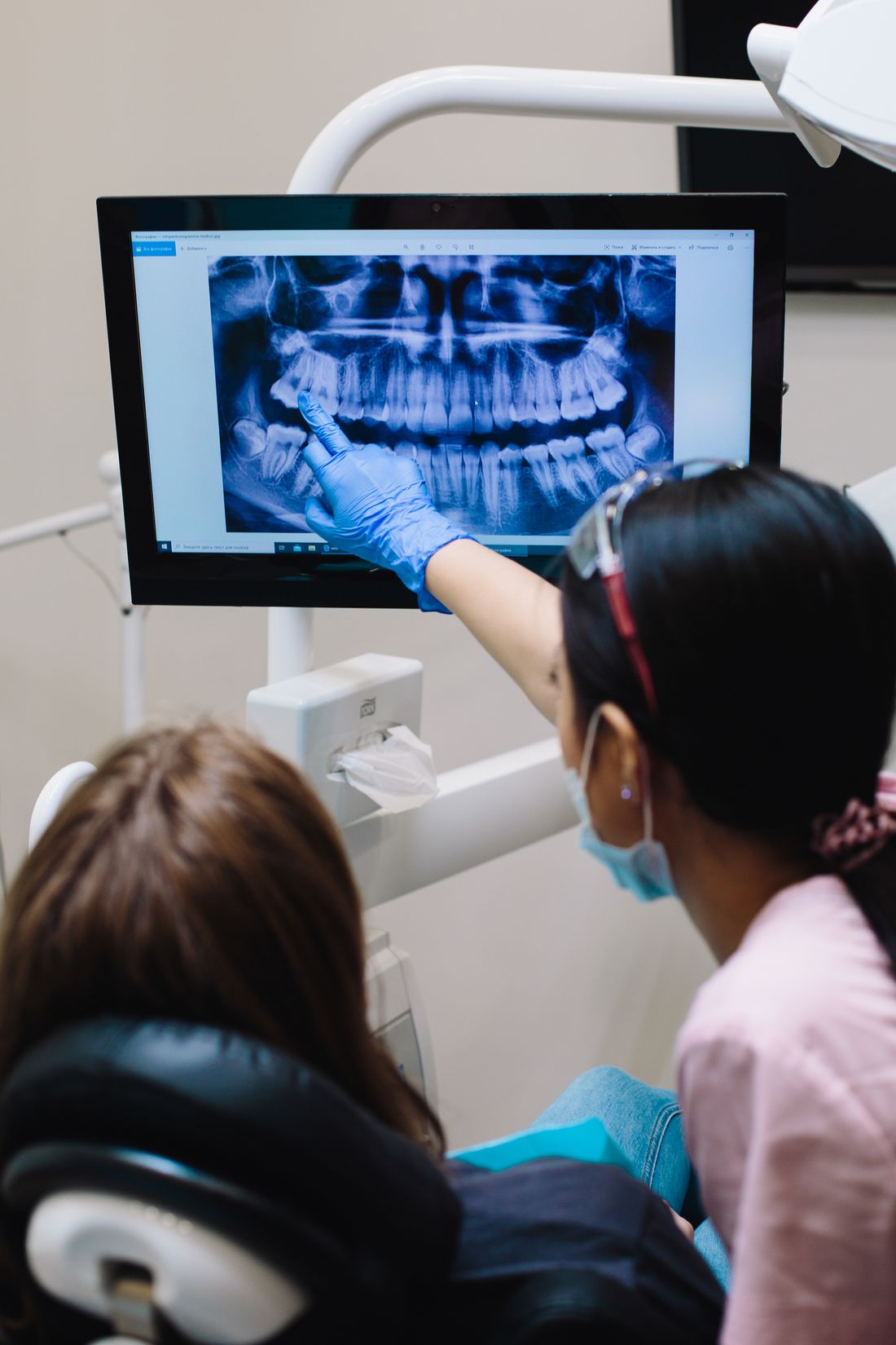
What does a dental cyst look like on an X-ray? Learn how cysts appear as dark, rounded areas, how types differ, and why a dentist must confirm.
Reviewed by the toothcheck Dental Team Independent dentist providing online second opinions.
Hearing that your X-ray shows a "cyst" or a "lesion" is unsettling, mostly because the word sounds ominous and nobody explained what they were pointing at. In reality, a dental (odontogenic) cyst is a fluid-filled sac that forms in the jawbone, usually from the remnants of tissue involved in tooth development or from long-standing inflammation. The great majority are benign. They are, however, worth taking seriously, because they can keep growing quietly.
This guide explains what cysts look like on a dental X-ray, why they look that way, how the common types differ, and where the honest limits of a 2D film lie. It is educational, not a way to diagnose your own image: the same dark spot can mean several different things, and telling them apart is a professional judgment.
Quick Answer
On a dental X-ray, a cyst typically appears as a well-defined, rounded or oval dark area (radiolucent) in the jawbone, often outlined by a thin white border. Its location points to the type: at the root tip of a tooth with a dead nerve suggests a radicular cyst, and around the crown of an unerupted tooth suggests a dentigerous cyst. However, small cysts, granulomas and abscesses can look alike on a 2D film, so the image alone cannot confirm the diagnosis.
Why a Cyst Looks Dark
X-rays pass easily through soft tissue and fluid but are absorbed by dense structures such as enamel, dentin and bone. Healthy bone appears in shades of white and gray because it is mineralized. A cyst is a cavity filled with fluid or soft material, so it absorbs far fewer X-rays and shows up as a darker (radiolucent) region where bone should be.
The thin white rim sometimes seen around a cyst is a layer of bone (a corticated margin) that the body lays down around a slowly expanding lesion. A smooth, sharply outlined edge generally reflects slow, contained growth. An ill-defined, ragged edge is more typical of faster-moving processes such as an active infection, which is one reason the border is one of the first things a dentist studies.
The Common Types and Where They Appear
The location of a dark area relative to the teeth is often the biggest clue. The table below summarizes the most common odontogenic cysts. These are typical patterns, not rules; individual cases vary.
| Cyst type | Typical location | Typical X-ray appearance | Usual associated tooth |
|---|---|---|---|
| Radicular (periapical) cyst | At the root tip of a tooth | Rounded dark area at the apex, often with a thin white border, continuous with the root outline | Non-vital (dead nerve) tooth, often with deep decay or a large old filling |
| Dentigerous cyst | Around the crown of an unerupted tooth | Dark area attached at the neck of the tooth, surrounding the crown like a halo | Impacted tooth, commonly a wisdom tooth or canine |
| Residual cyst | In the site of a previously extracted tooth | Rounded dark area in the bone where the root once was | Missing tooth (an old radicular cyst left behind) |
| Odontogenic keratocyst | Often the back of the lower jaw, can extend along the bone | Dark area, sometimes multi-lobed, tends to grow along the jaw rather than swell it | May or may not involve a tooth |
Radicular (Periapical) Cysts
The radicular cyst is the most common cyst of the jaws. It develops at the tip of a tooth whose nerve has died, usually after deep decay, trauma or a failed old restoration. Bacteria and breakdown products leak out of the root tip and the body responds with chronic inflammation, producing a granuloma (a mass of inflamed tissue). In some granulomas, leftover epithelial cells in the area proliferate and form a fluid-filled lining, which becomes a cyst.
On film, the dark area is centered on the root tip, and the thin white line (lamina dura) that normally outlines the root in the bone is lost there. These cysts are frequently painless. They are generally found on routine X-rays or when the tooth flares up.
Dentigerous Cysts
A dentigerous cyst forms around the crown of a tooth that has not come through the gum, attached where the enamel meets the root. It is the second most common type and most often involves lower wisdom teeth and upper canines. The X-ray shows a smooth, dark, rounded space around the crown, which can look like an enlarged "follicle," the normal soft-tissue sac around a developing tooth.
That is a genuine diagnostic subtlety: a small amount of dark space around an unerupted crown is normal, and a cyst is suspected when the space is clearly widened, often more than a few millimeters, or when the tooth is being pushed out of position. If you are looking at an impacted wisdom tooth, see our guide on what an impacted wisdom tooth looks like on an X-ray.
Residual Cysts and Keratocysts
A residual cyst is simply a radicular cyst that was left behind when its tooth was removed, so it appears as a dark area in an extraction site. Odontogenic keratocysts are less common but notable because they behave differently: they tend to grow lengthwise through the bone, may recur after removal, and are recognized by their lining under the microscope. Their appearance on a 2D film overlaps with other cysts, which is one reason tissue examination matters for diagnosis.
Cyst, Granuloma or Abscess? Why It Is Hard on a 2D Film
This is the part that surprises most patients. A small cyst, a granuloma and an abscess at a root tip can all appear as a dark area at the apex. A 2D film also flattens a three-dimensional structure into a single plane, so overlapping bone and teeth can hide or mimic features.
Clues that help a dentist distinguish them include:
- Size: radicular lesions larger than roughly 1 centimeter are more often cysts than granulomas, though this is a tendency, not a rule.
- Border: a smooth, corticated border suggests slow growth, while a diffuse, fading edge suggests active infection.
- Tooth vitality: a cold or electric test showing the tooth has no living nerve points toward a radicular origin.
- Symptoms: swelling, pain on biting, a gum bump or drainage point toward an abscess or infected lesion.
- Change over time: comparing with older films shows whether the area is growing.
For the abscess side of this comparison, see what a tooth abscess looks like on an X-ray. Even with all of these clues, a definitive diagnosis of a jaw lesion sometimes requires a biopsy, where the removed tissue is examined under a microscope. A small number of other conditions, including some tumors and non-dental bone lesions, can resemble a cyst on film, which is exactly why a dark area should not be ignored or self-diagnosed.
Do Cysts Need Treatment?
Most cysts that are confirmed or strongly suspected are treated, because they do not shrink on their own and can slowly enlarge, displace or loosen teeth, thin the jawbone, or become infected. The approach depends on type and size:
- Radicular cyst: often managed by root canal treatment of the source tooth, sometimes with a small surgical procedure at the root tip, or by removing the tooth. Some small lesions heal substantially after the infection source is treated.
- Dentigerous cyst: usually removed together with the impacted tooth, or the tooth is guided into position if it is worth saving.
- Larger cysts: may be decompressed first (drained to let the cyst shrink) and then removed (enucleation).
- Tissue testing: removed material is commonly sent to a laboratory to confirm what it was.
Because treatment can range from monitoring or a root canal to oral surgery, it is reasonable to understand why a specific plan was recommended.
When 2D Is Not Enough
A standard periapical or panoramic X-ray gives a flat view. For larger lesions, or when the exact extent, relationship to nerves or neighboring roots matters for surgical planning, in-person assessment often includes cone beam CT (CBCT), which shows the jaw in three dimensions. Toothcheck does not interpret CBCT. If you want to understand which image suits which question, see which dental images are used for which treatment, and for a general primer, reading dental X-rays: what you can see.
Getting a Second Opinion on a Cyst Finding
If a cyst or lesion has been found and a procedure has been proposed, a written second opinion can be useful. A licensed dentist reviews your uploaded X-ray and the treatment plan, and comments on whether the findings and the recommended steps appear consistent with the image, what alternatives might exist, and what questions to bring back to your treating dentist or specialist. Our service is an online dental second opinion for 59 dollars with a written response in about 72 hours. It works from 2D X-rays and does not replace an in-person exam, a CBCT interpretation or a biopsy.
For general background on oral health conditions, the American Dental Association and the National Institute of Dental and Craniofacial Research provide patient information.
FAQ
What does a dental cyst look like on an X-ray? Typically a well-defined, rounded or oval dark area in the jawbone, often outlined by a thin white border. A radicular cyst sits at the root tip of a tooth with a dead nerve, while a dentigerous cyst surrounds the crown of an unerupted tooth. Appearance alone is not a diagnosis.
Can a dentist tell a cyst from an abscess on an X-ray? Not always. On a 2D film a small cyst, a granuloma and an abscess can all look like a dark area at a root tip. Size, shape, border, tooth vitality and symptoms all help, and a biopsy sometimes provides the definitive answer.
Are dental cysts dangerous? Most are benign, but they can slowly enlarge, displace teeth, weaken the jawbone and become infected. A few lesions that resemble cysts are more significant, so a cyst-like finding should be evaluated rather than ignored, without cause for panic.
Does a dental cyst always cause pain? No. Many are found by chance on routine X-rays because they grow slowly and cause no symptoms. Pain, swelling, a bad taste or a gum bump usually signals infection or a larger lesion.
How are dental cysts treated? It depends on the type and size. A radicular cyst may resolve after root canal treatment or extraction of the involved tooth. Others are removed surgically or decompressed first, and tissue is often sent to a lab for confirmation.
Can toothcheck diagnose a cyst from my X-ray? No. A licensed dentist can review your 2D X-ray and treatment plan and give a written second opinion, but we do not interpret CBCT scans and cannot replace an in-person exam or biopsy.
Final Thoughts
A dark, rounded area on an X-ray is a reason to follow up, not a reason to panic. Most dental cysts are benign and treatable, but the image alone cannot separate a cyst from a granuloma, an abscess or, rarely, something more significant. This article is educational and is not a diagnosis or a substitute for an in-person examination. If a cyst has been mentioned and you want a clear, independent review of the X-ray and the plan, get an online dental second opinion.
Last medically reviewed: October 2026