What Does a Tooth Infection Look Like on an X-Ray?
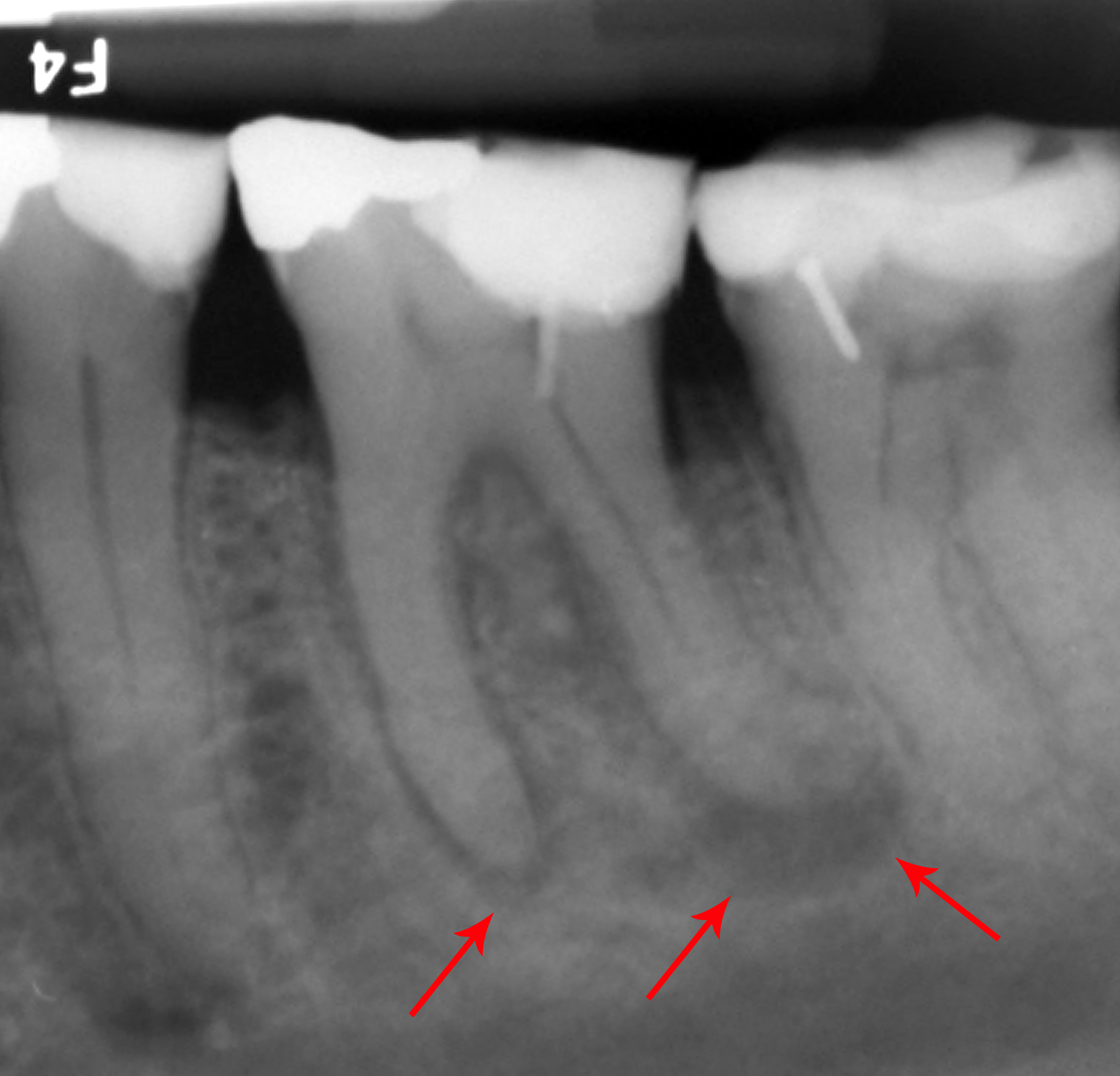
A dentist-validated guide to spotting a tooth infection on a dental X-ray: the dark area at the root tip, early signs, what mimics it, and when it is urgent.
Validated by our dental expert team Independent dentists providing online second opinions.
Your dentist points near the bottom of a tooth root, says the word "infection," and suddenly you are staring at a grey image trying to see what they see. The good news is that a tooth infection has a fairly recognizable signature on an X-ray once someone shows you where to look. This guide walks through exactly what it looks like, how to spot it on your own film, and, just as importantly, what a dark spot does not always mean.
Quick Answer
A tooth infection usually shows up on an X-ray as a dark (radiolucent) area at the tip of a tooth root, called a periapical radiolucency. It forms when the nerve inside the tooth has died or become infected and bacteria leak out of the root end into the surrounding bone, which the infection gradually dissolves, and less-dense areas read dark on an X-ray. Earlier signs are subtler: a widening of the thin dark line around the root and loss of the bright white outline beside it. A dark spot at a root tip is a finding, not an automatic emergency, however, because some are actively painful while others are long-standing and stable. Your symptoms, the tooth's history, and an in-person exam decide how urgent it is. If a finding like this turns up on your plan, an online dental second opinion can explain what it appears to show.
Worried that dark spot on your X-ray is an infection? You cannot be sure from the image alone. An independent, licensed dentist will review your actual X-ray and treatment plan in writing within 72 hours for a flat $59 — get an X-ray second opinion or start an online second opinion.
Where a tooth infection shows up: the root tip
To read the sign, it helps to know the story behind it. Inside every tooth is a living pulp (the nerve and blood supply). When deep decay, a crack, or trauma lets bacteria reach the pulp, the nerve can become inflamed and eventually die. Once the pulp dies, the infection travels down the inside of the tooth and exits through the tiny opening at the tip of the root, into the bone around it. That is why a tooth infection almost always shows up at the apex (the root tip), not up in the crown. The medical term "periapical" simply means "around the tip of the root."
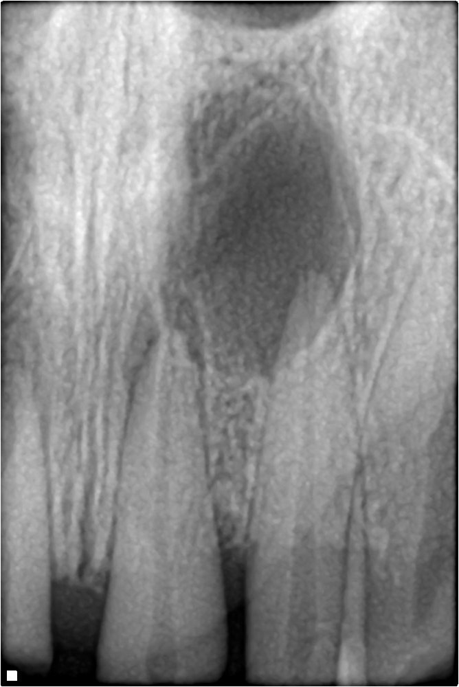
The telltale sign: a dark circle at the root tip
The classic, mature sign of a tooth infection is a dark, often rounded area hugging the tip of the root, like a shadow where solid bone should be. Bone is dense and reads as a fairly even grey-white on an X-ray; when infection dissolves a pocket of that bone and replaces it with pus and inflamed tissue, that area becomes less dense and therefore darker on the image.
A few features a dentist reads from it:
- Location. Centered on the very tip of the root, not along the side of the crown.
- Definition. A well-defined, clearly outlined dark circle often suggests a long-standing, chronic lesion. A fuzzier, more diffuse dark area can suggest a more active or acute process.
- Size. Larger areas mean more bone has been lost, though size alone does not decide urgency.
An abscess versus a long-standing (chronic) lesion
Not every root-tip infection behaves the same way, and the X-ray often hints at which kind you are looking at.
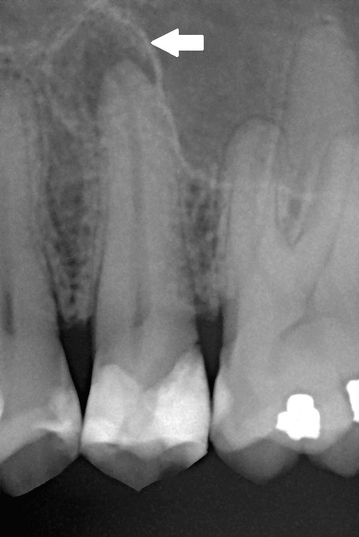
An acute periapical abscess is an active, often painful infection. It may come with swelling, throbbing, pain on biting, or a "pimple" on the gum (a draining fistula), and on an X-ray the dark area can look less sharply defined. A chronic lesion (sometimes called chronic apical periodontitis, and often a granuloma or cyst on examination) has smoldered for a long time; it is frequently well-outlined and round, and it can be completely painless, which is why some infections are discovered only on a routine X-ray. Whether a lesion is a granuloma or a cyst usually cannot be told from the image alone. The plain-English versions of these terms are collected in our guide to common dental diagnoses explained.
Why infection reads dark: the radiolucent rule
If you remember one principle, make it this: dense things read white, and less-dense things read dark. Metal fillings, crowns, and root fillings block X-rays completely and appear bright white (radiopaque). Bone is moderately dense and reads grey-white. Infection, pus, decay, and air are less dense than bone, so they read dark (radiolucent). A tooth infection is essentially a dark hole eaten into bright bone, which is exactly why it stands out once you know to look for it. For a fuller tour of the light and dark areas, see the dental X-ray interpretation guide for patients.
The early and subtle signs most people miss
A brand-new infection does not always show a big dark circle yet, because it takes time for enough bone to be lost to be visible. Before the obvious stage, a dentist may notice:
- A widened dark line around the root. A healthy tooth has a thin, even dark line (the periodontal ligament space) around its root. Early infection can widen that line at the tip.
- Loss of the bright white outline. A thin white line (the lamina dura) normally traces the root socket. When it fades or disappears at the apex, that can be an early clue.
- A trail from a deep cavity. A large dark area of decay reaching the pulp chamber, connected to a shadow at the root tip, tells a consistent story of how the infection got there.
Because these early signs are subtle and easy to misread, they are a common reason to get a second set of eyes on a film. Our page on understanding your dental X-ray report and chart notes decodes the shorthand a dentist may write for them.
Tips for spotting it on your own X-ray
1. Follow each root down to its tip. Infection lives at the apex, so let your eye travel to the very end of the root. 2. Compare with the neighbors. Healthy root tips look similar to each other. A dark pocket at one tip that its neighbors do not have is the thing to notice. 3. Look for a break in the bone pattern. You are hunting for a dark pocket where the bone should be an even grey. 4. Check that tooth for a reason. A deep filling, a crown, or obvious decay on the same tooth is a plausible reason the nerve died, which makes an infection more likely. 5. Confirm which tooth it is. Match the spot to a tooth number so you can discuss it precisely; our guide on how to identify tooth numbers on a dental X-ray shows you how.
What looks like an infection but is not
This is where caution matters, because several normal, harmless things can imitate a dark spot at a root:
- Normal anatomy. The mental foramen (a normal opening near the lower premolars), the maxillary sinus above upper back teeth, the incisive foramen behind the upper front teeth, and small nutrient canals can all appear as dark areas near roots.
- A healed scar. After a successful root canal, a small dark area can persist as scar tissue even though the infection is gone.
- Overlap. X-rays are flat pictures of 3D anatomy, so structures can overlap and create a shadow that is not really at the root tip.
Because of these look-alikes, a responsible reading pairs the image with vitality testing and your symptoms rather than diagnosing from a shadow alone.
What an X-ray cannot tell you by itself
An X-ray is powerful but limited. It cannot feel your pain, it cannot always distinguish a granuloma from a cyst, and it cannot by itself confirm whether a nerve is alive (that needs a cold or electric pulp test). It is also a 2D snapshot, so a small or early lesion may not be visible yet. And a dark area that is real can still be stable and long-standing rather than an emergency. In other words, the film raises the question; your dentist's exam answers it.
Why it matters and what to do
A genuine, active tooth infection does not resolve on its own, and left alone it can grow, cause pain, and occasionally spread, so it is worth taking seriously. The usual treatments are a root canal to clean out the infected inside of the tooth, or extraction if the tooth cannot be saved; antibiotics may calm a flare-up but do not cure the source. If you want to know whether the finding truly points to treatment, our guides on the signs you need a root canal and whether you really need a root canal walk through it, and what to do about a tooth abscess covers the urgent end of the spectrum. The American Association of Endodontists is a reliable patient resource on root-tip infections.
FAQ
What does a tooth infection look like on an X-ray? It typically appears as a dark (radiolucent) area at the tip of a tooth root, where infection has dissolved some of the surrounding bone. Earlier signs are subtler, such as a widened dark line around the root tip and loss of the thin white outline beside it.
Does a dark spot on my X-ray always mean an infection? No. Normal structures like the mental foramen or the maxillary sinus, a healed scar after a root canal, or simple overlap of anatomy can all mimic a dark spot. That is why the image is read alongside your symptoms and a clinical exam rather than on its own.
Can a tooth be infected but not show up on an X-ray? Yes. It takes time and enough bone loss for an infection to become visible, so an early infection may not show yet. A dentist may rely on symptoms and nerve-vitality testing when the X-ray still looks normal.
Is a dark area at the root tip an emergency? Not always. Some root-tip lesions are active and painful, while others are chronic, stable, and even painless for years. Whether it is urgent depends on your symptoms, signs of spreading infection, and whether it is growing over time.
What is the difference between an abscess, a granuloma, and a cyst on the image? All three can look like a dark area at the root tip, and the X-ray alone usually cannot tell them apart. An acute abscess tends to be more diffuse and symptomatic, while chronic granulomas and cysts are often well-defined and round; distinguishing them may need further testing.
How is a tooth infection treated once it is seen? The common options are a root canal, which cleans out the infected inside of the tooth, or extraction when the tooth cannot be saved. Antibiotics can settle a flare-up but do not remove the source, so they are not a standalone cure.
Final Advice
A tooth infection has a clear signature once you know it lives at the root tip and reads as a dark pocket in the bone. But the same picture can be mimicked by normal anatomy or a healed scar, and a real lesion can be either an emergency or a stable, long-standing finding. That gap between "there is a shadow" and "here is what it means for you" is exactly where a calm, plain-English explanation helps.
This guide was reviewed and validated by our dental expert team before publishing. If a dark spot or the word "infection" has turned up on your X-ray or treatment plan, you can upload your dental X-rays for a second opinion or ask for a full treatment plan review, and a licensed dentist will explain what it appears to show, usually in under 72 hours.
Image credits: Periapical abscess radiograph by Coronation Dental Specialty Group via Wikimedia Commons (CC BY-SA 3.0); periapical radiolucency image by Shaimaa Abdellatif via Wikimedia Commons (CC BY-SA 4.0); chronic apical periodontitis image by Michele Gardini via Wikimedia Commons (CC BY-SA 3.0). Used as educational examples — they do not depict toothcheck patients.
Last medically reviewed: July 2026